Alaskan Husky Encephalopathy is a serious canine neurodegenerative disorder that affects Alaskan Huskies, a breed of sled dog.
It's estimated that the disorder affects up to 20% of the Alaskan Husky population.
Symptoms of the disorder can include seizures, tremors, and loss of coordination.
Affected dogs often exhibit a characteristic "shaking" or "trembling" of their head and body.
The disorder is typically diagnosed between the ages of 2-6 years, with males being more susceptible than females.
Unfortunately, there is no known cure for Alaskan Husky Encephalopathy.
Introduction
Alaskan Husky Encephalopathy is a rare and serious condition that affects a specific breed of dog. It's a genetic disorder that can be devastating for dog owners.
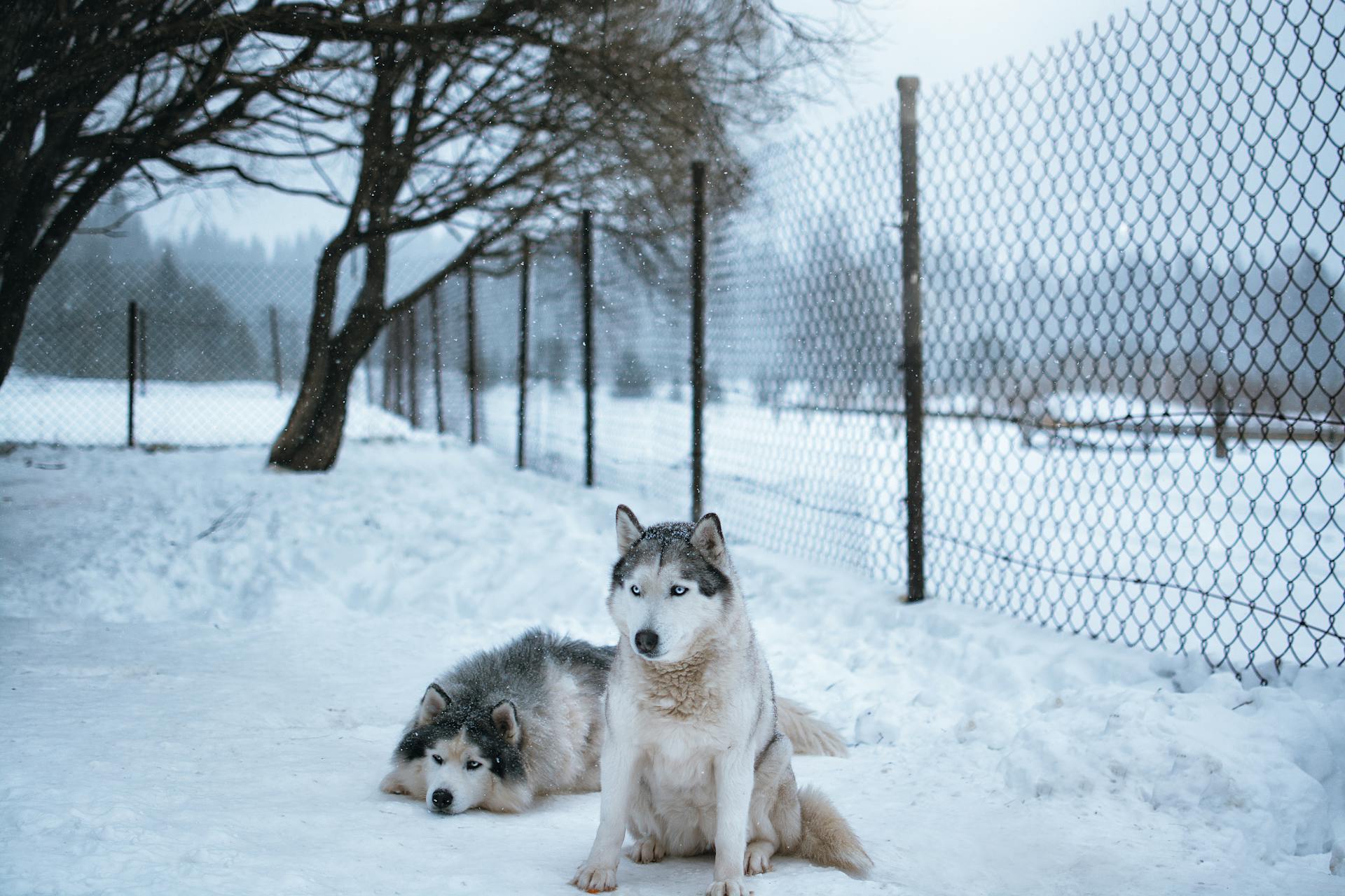
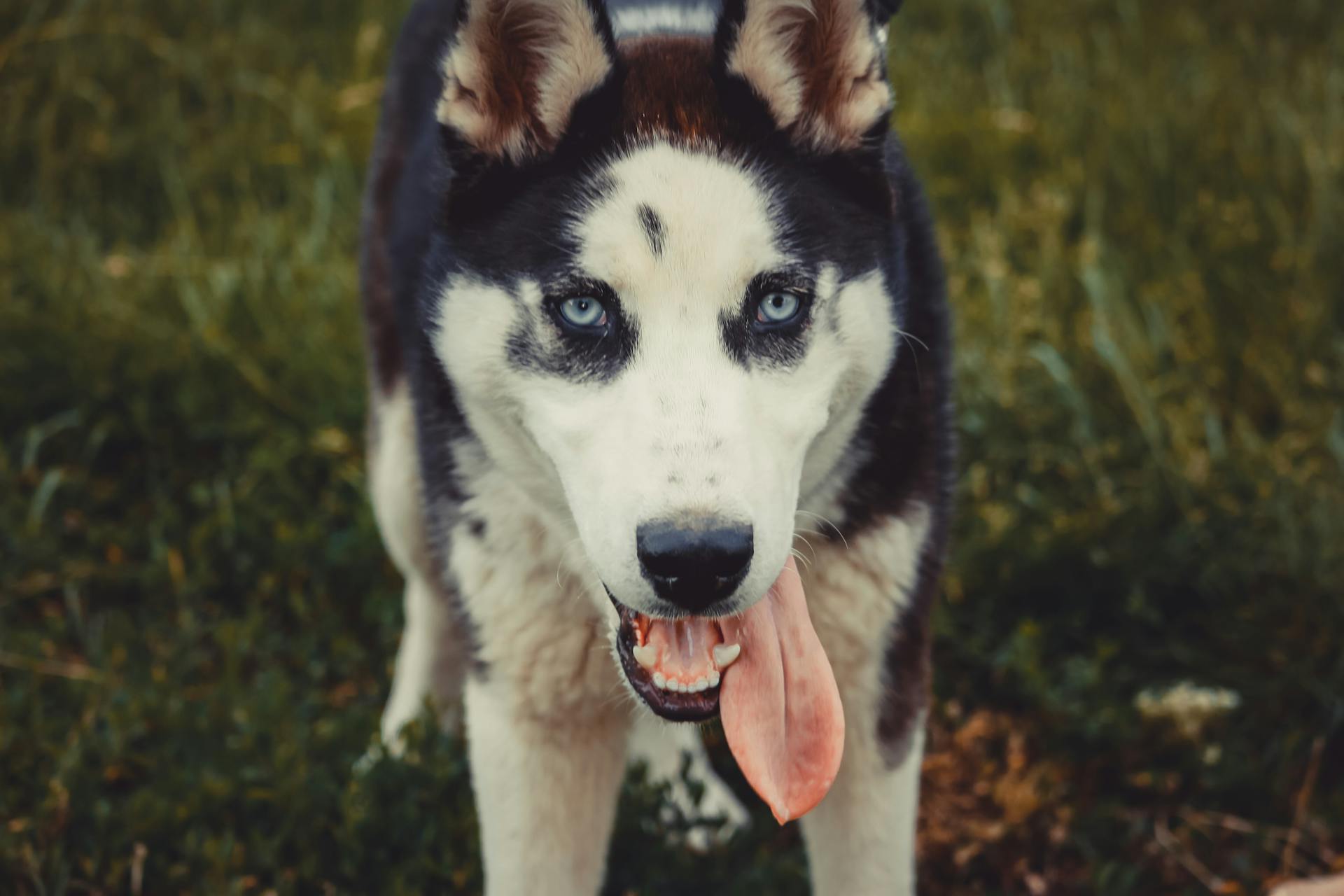
Alaskan Huskies are a popular breed known for their intelligence, athleticism, and friendly nature. They were originally bred to pull sleds in the Arctic.
The condition is caused by a mutation in the gene that codes for a protein called SLC3A1. This mutation can lead to a buildup of amino acids in the brain, causing damage and inflammation.
Alaskan Huskies can be prone to this condition due to their genetic makeup.
Causes and Genetics
Alaskan Husky Encephalopathy is caused by a mutation in the SLC19A3 gene, which affects the production of a protein essential for brain function. This mutation leads to a frameshift and premature peptide termination, resulting in a truncated protein.
The mutation was identified through sequencing of the SLC19A3 gene in dogs with AHE, and it was found to be homozygous in all 11 affected dogs. In contrast, 26 out of 41 unaffected Alaskan Husky dogs were homozygous wild type, while 15 were heterozygous carriers.
The mutation is not a polymorphism, as it was not identified in 187 dogs from 51 breeds. This suggests that the mutation is specific to the Alaskan Husky breed and is likely the primary cause of AHE.
Genetics and Characteristics
Genetics plays a significant role in determining an individual's susceptibility to certain conditions. Research has identified multiple genetic variants associated with an increased risk of developing a specific condition.
Genetic testing can help identify these variants, allowing for early intervention and potentially altering the course of the condition. This can be especially beneficial for individuals with a family history of the condition.
A combination of genetic and environmental factors contributes to the development of a condition. This can make it challenging to pinpoint a single cause, but understanding the genetic component can provide valuable insights.
Studies have shown that certain genetic variants can affect the functioning of specific organs or systems in the body. This can increase the risk of complications and make management more complex.
Genetic counseling can help individuals understand their genetic risk and make informed decisions about their health. It's essential to discuss genetic testing and counseling options with a healthcare professional.
SLC19A3 Sequencing
Sequencing of the SLC19A3 gene revealed some important changes in dogs with AHE.
Four changes were identified in dogs with AHE compared to unaffected dogs, including a 4 bp insertion and a SNP in exon 2.
This insertion and SNP were predicted to result in a frame shift and premature peptide termination, truncating 279 amino acids of the full-length protein.
A genotyping assay based on PCR product size was developed to test additional animals.
All 11 dogs with AHE were homozygous for the mutation, while 26 out of 41 unaffected dogs were homozygous wild type.
15 out of 41 unaffected dogs were determined to be heterozygous carriers of the mutation.
The mutant allele was not identified in a group of 187 dogs from 51 breeds, suggesting it's not a common polymorphism.
Expression levels of SLC19A3.1 were compared in cerebral cortex tissue from an affected dog and an unaffected beagle, and SLC19A3.1 expression was dramatically decreased in the affected dog.
Clinical Presentation
Alaskan Husky encephalopathy (AHE) is a serious condition that affects the brains of these dogs. The clinical presentation of AHE is characterized by typical clinical signs.
Dogs with AHE often exhibit generalized seizures, gait abnormalities, dysphagia, and ataxia. These symptoms can vary in severity and progression.

All 11 dogs with AHE included in one study showed typical clinical signs, including seizures, gait abnormalities, and ataxia. The symptoms were often static or progressed rapidly.
The age of onset for AHE can vary, but in one study, the affected dogs were euthanized or showed significant neurological deficits at an average age of 2-3 years.
Diagnostic Methods
Diagnostic tests were conducted to rule out other potential causes of the Alaskan Husky encephalopathy. No abnormalities were noted on the complete blood count (CBC).
The cerebrospinal fluid (CSF) and serum biochemistry analysis also showed no indication of any underlying health issues. In fact, the analysis revealed no abnormalities in any dog.
The evaluation of plasma or CSF lactate or pyruvate levels, or in the lactate to pyruvate ratios, showed no indication of a mitochondrial disorder.
Biochemical Assays in Blood and CSF
Biochemical assays in blood and cerebrospinal fluid (CSF) are a crucial part of the diagnostic process.
No abnormalities were noted on the CBC, CSF, and serum biochemistry analysis in any dog.
These tests checked for various factors, including lactate and pyruvate levels in plasma and CSF.
The lactate to pyruvate ratios were also evaluated, but there was no indication of a mitochondrial disorder.
This suggests that the biochemical assays in blood and CSF are a reliable way to rule out certain conditions.
In fact, the tests were thorough enough to detect any abnormalities, which were not found in any of the dogs.
RT-PCR
RT-PCR is a diagnostic method used to compare the expression of specific genes in different tissues. This method is semiquantitative, meaning it provides a relative measure of gene expression.
The RT-PCR technique involves making cDNA from various tissues, including spleen, skin, cerebellum, thymus, testis, spinal cord, heart, muscle, cerebral cortex, kidney, and liver.
A housekeeping gene, GAPDH, is used as a baseline to ensure equal concentrations and quality of the samples for expression comparison. This is crucial for accurate results.
RT-PCR can be performed using RNA extracted from specific tissues, such as the cerebral cortex of an affected and an unaffected non-AH dog. This allows for direct comparison of gene expression between the two samples.
Pathology and Research
Seven dogs were euthanized and examined for neuropathological lesions characteristic of Alaskan Husky Encephalopathy (AHE).
These lesions were found in the thalamus, claustrum, cerebral cortex, putamen, caudate nucleus, globus pallidus, pons, and medulla oblongata, and were bilaterally symmetrical.
Microscopically, lesions included foci of polioencephalomalacia and necrosis with incipient malacia in the deep gray and underlying white matter of the cerebral cortex.
Similar but milder malacic lesions were also found in the deep in the midline of the ventral lobes of the vermis.
In some cases, muscle tissue from affected dogs showed deposits of succinic dehydrogenase (SDH) positive material, most consistent with mild mitochondrial proliferation.
Ultrastructurally, enlarged megamitochondria were observed in affected dogs, with abnormal glycogen deposits inside the mitochondria.
Neuropathology Findings
In neuropathology, lesions characteristic of AHE were found in all seven dogs studied. These lesions were bilaterally symmetrical and located in various areas of the brain.
The thalamus showed grossly visible cystic encephalomalacia. This is a type of brain damage that can occur due to various causes.
Polioencephalomalacia, a condition where brain tissue dies, was also found in the claustrum. This area is responsible for processing sensory information.
Necrosis, or cell death, was observed in the deep gray and underlying white matter of the cerebral cortex, as well as in the putamen, caudate nucleus, globus pallidus, pons, and medulla oblongata.
Similar but milder lesions were found in the deep midline of the ventral lobes of the vermis.
In two dogs, muscle tissue was evaluated and found to be normal in one, but showed mild mitochondrial proliferation in the other. This was indicated by deposits of succinic dehydrogenase and cytochrome c oxidase positive material.
Ultrastructural lesions were observed in the skeletal muscle of two affected dogs, including enlarged megamitochondria. These megamitochondria were 2-9 μm in diameter, compared to 0.5 μm in a normal dog.
Genome-Wide Association Study
A Genome-Wide Association Study was conducted on 8 affected dogs and 20 unaffected but related controls.
The study used 114,613 SNPs after pruning, and a genomic inflation factor of 1 indicated little population stratification.
A cluster of SNPs on CFA 25 showed raw p values 56 times lower than the next best associated SNP on CFA 30.
Permutation testing did not give genome-wide significance, but the strong signal from many SNPs from one discrete location warranted further investigation.
A region of complete homozygosity was identified in all affected dogs and never in the 20 unaffected dogs.
This region extended 3.5 Mb between 41074763 and 44542154 on CFA 25.
The best associated SNP was located at 43980115 Mb on CFA 25, with a p value of 6.59×10.
Discussion
The field of pathology and research is a rapidly evolving one, with new discoveries being made every day.
One of the key areas of focus in pathology research is the study of biomarkers, which can help diagnose diseases more accurately and earlier than ever before.
Biomarkers can be found in various bodily fluids, including blood and urine, and can be used to track the progression of a disease or the effectiveness of a treatment.
Researchers have identified several biomarkers that are associated with specific diseases, such as cancer and cardiovascular disease.
The use of biomarkers in pathology research has led to the development of new diagnostic tests and treatments.
The study of biomarkers has also led to a better understanding of the underlying causes of diseases, which can inform the development of new treatments and therapies.
Pathology research has also led to the development of new technologies, such as advanced imaging techniques and robotic surgery.
These technologies have improved the accuracy and speed of diagnosis, as well as the effectiveness of treatments.
The collaboration between researchers, clinicians, and industry partners has been crucial in the advancement of pathology research.
This collaboration has led to the development of new products and services that can improve patient outcomes.
Affected Breed
The Alaskan Husky breed is particularly affected by Alaskan Husky Encephalopathy (AHE).
Dogs with AHE exhibit clinical signs such as generalized seizures and gait abnormalities.
These symptoms can also include dysphagia and ataxia.
AHE is characterized by specific neuroimaging and neuropathological lesions.
Bilaterally symmetrical, cavitatory malacic lesions are found in the thalamus.
Smaller malacic lesions are also present in the putamen, claustrum, junctional grey and white matter in the cerebral cortex, brainstem, and midline cerebellar vermis.
These lesions are hyperintense on T2W MR images.
Frequently Asked Questions
What age do dogs get Alaskan husky encephalopathy?
Dogs typically develop Alaskan husky encephalopathy between 6 months to 3 years of age, with symptoms either appearing suddenly or progressing gradually over time.
Sources
- https://pubmed.ncbi.nlm.nih.gov/10912920/
- https://www.pawprintgenetics.com/products/tests/details/124/
- http://www.animalabs.com/shop/dogs/alaskan-husky-encephalopathy-ahe/
- https://journals.plos.org/plosone/article
- https://escholarship.org/uc/item/10q2v8mm
- https://journals.plos.org/plosone/article/figures
Featured Images: pexels.com