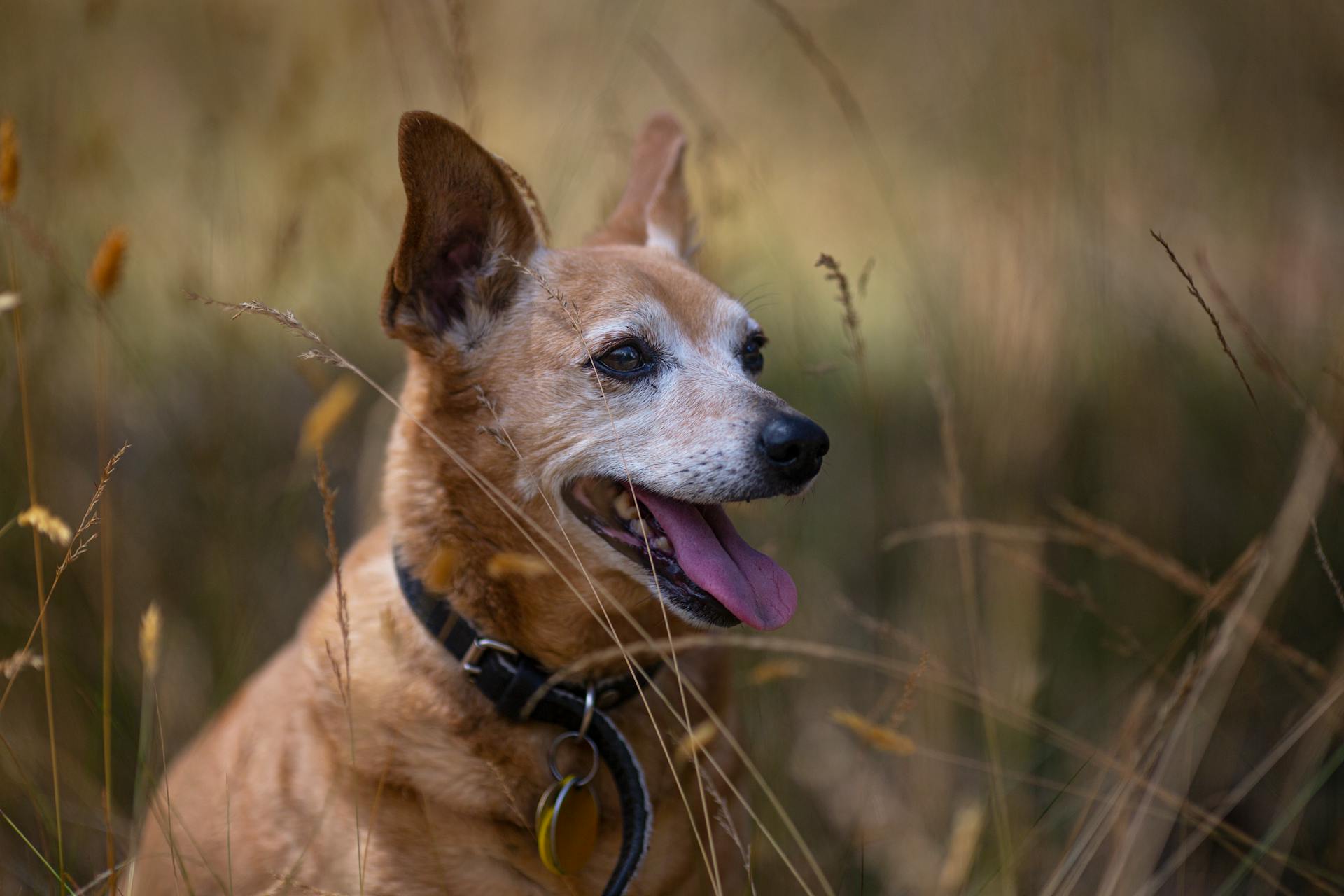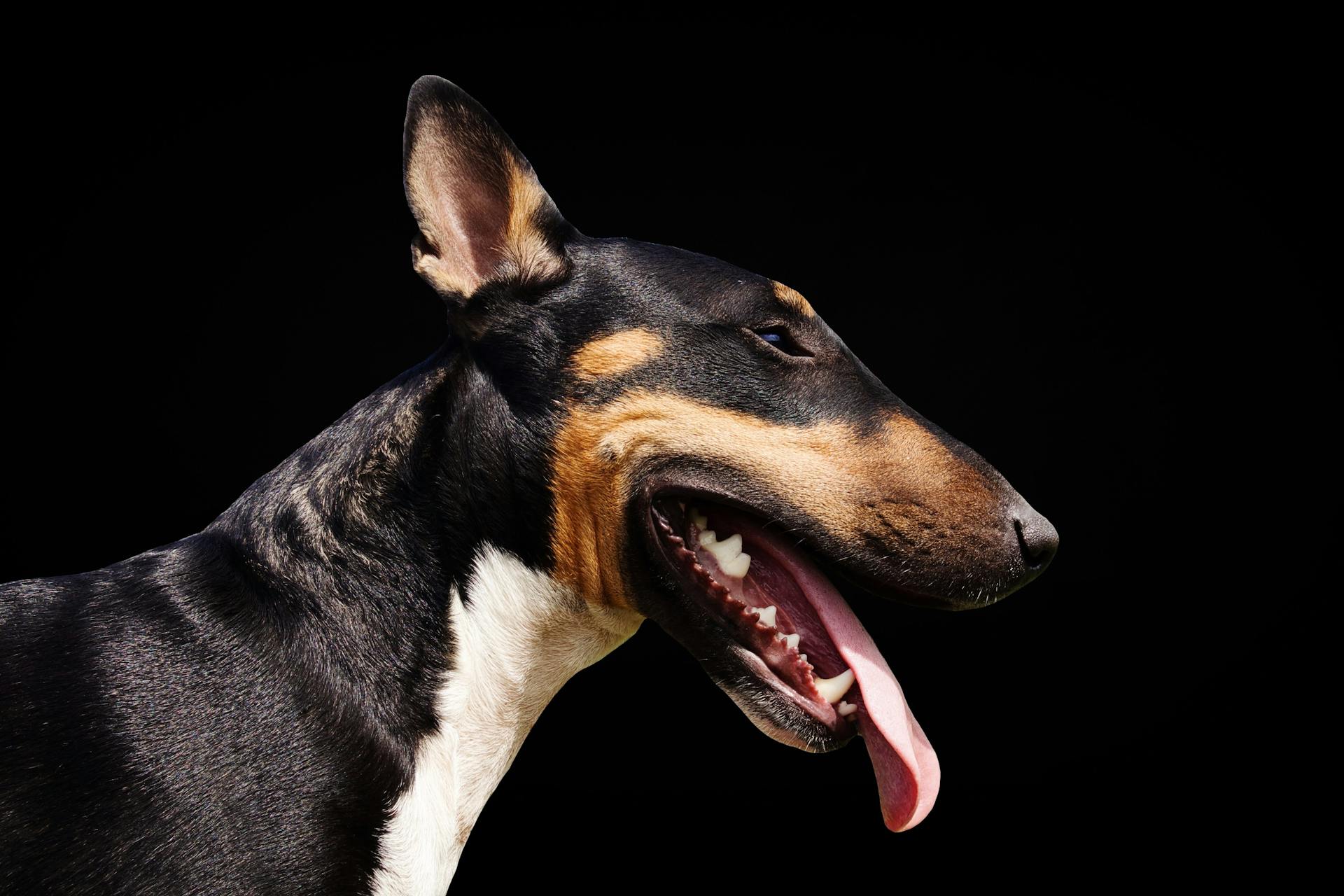
The canine brain is a remarkable organ that plays a crucial role in shaping our furry friends' behavior.
Dogs have a highly developed sense of smell, thanks to their large olfactory bulb, which is responsible for processing smells.
Their brain-to-body mass ratio is relatively small compared to humans, but their brain is still incredibly efficient at processing information.
In fact, studies have shown that dogs can learn and remember hundreds of words, including names, actions, and objects.
Dogs also have a unique way of processing emotions, with research suggesting that they can experience emotions similar to humans, such as joy and excitement.
A fresh viewpoint: Canine Ear Canal Anatomy
Canine Brain Anatomy
The canine brain has a unique anatomy that has been studied extensively in neuroscience experiments. The brain's surface can be reconstructed using a modified version of the FreeSurfer toolkit, which allows for accurate identification of gray and white matter boundaries.
The high-resolution template of the canine brain is created by processing images using the FreeSurfer toolkit, which automatically segments the brain into different tissues. However, some regions may require manual intervention to demarcate the boundary due to partial volume effects.
The gray and white matter intensity differences in the canine brain are very large, making the corresponding tissue segmentation generally accurate. The white matter surface can be inflated using standard tools available in FreeSurfer, creating a smoothed surface that can be used for further analysis.
Suggestion: Water on the Brain in Dogs
The Canine Experience
Dogs have been serving as experimental models in neuroscience for several centuries, with some of the earliest evidence for specific brain function localization coming from experiments on dogs by Gustav Fritsch and Eduard Hitzig.
These early experiments involved electrically stimulating small regions of the exposed cortex in awake animals, leading to significant discoveries about brain function.
Sir David Ferrier used similar techniques to identify multiple cortical areas related to the precise control of movement, which he later translated to map the "eloquent" cortex of patients with tumors undergoing neurosurgical procedures.
The dog has also been a valuable model for studying inherited retinal disease, with gene therapeutic treatment of these disorders being a major area of research.
Dogs suffer from age-related cognitive dysfunction, which resembles human Alzheimer's Disease, making them an important model system for studying this condition.
The dog's brain has been studied using functional and anatomical magnetic resonance imaging, with early studies showing that visual stimulation in the anesthetized dog can produce measurable changes in blood oxygen level dependent (BOLD) fMRI signal from the canine visual cortex.
A recent fMRI study examined the neural correlates of reward mechanisms in the awake dog, demonstrating the complexity of canine brain function.
A different take: Rabbit Brain Test
Smell
Dogs have an incredible sense of smell, thanks to their large olfactory cortex, which is dedicated to processing smells.
They have roughly forty times more smell-sensitive receptors than humans, with some breeds like bloodhounds having up to 300 million receptors.
This means dogs can detect scents that are too faint for us to smell, and they can even follow a scent trail that's hours old.
In fact, dogs can detect smells that are too subtle for humans to detect, making them invaluable for tasks like search and rescue.
Discover more: Dogs Anatomy Male
Imaging Techniques
Magnetic resonance imaging (MRI) is a crucial technique in studying canine brain anatomy. MRI was used to create in-vivo templates of dog brains.
Dogs were anesthetized and paralyzed during MRI acquisition to ensure accurate and consistent images. A 3 Tesla Siemens Trio scanner was used to acquire 15-minute MPRAGE images with 1 mm isotropic resolution.
The MRI protocol involved a transmit-receive, quadrature volume head coil and a 6-parameter realignment with least-squares minimization. This process allowed for the creation of high-quality, in-vivo images of dog brains.
In-vivo T1-weighted images were obtained from seven dogs, and the two MPRAGE images were averaged to create a single image. This averaging process helped to reduce noise and improve image quality.
Dogs imaged for template creation were premedicated with dexmedetomidine and induced to general anesthesia with propofol. They were maintained under anesthesia with inhalant isoflurane and oxygen with a dexmedetomidine continuous rate infusion.
A 3.0T General Electric Discovery MR750 scanner was used to perform MRI examinations, operating at 50mT/m amplitude and 200T/m/s slew-rate. This scanner was equipped with a 16-channel medium flex radio-frequency coil.
A high-resolution T1-weighted 3D inversion-recovery fast spoiled gradient echo sequence was performed in each subject with isotropic voxels of 0.5 mm. This sequence helped to create detailed images of dog brains.
Dogs imaged for skull shape compatibility validation were premedicated with methadone and induced to general anesthesia with propofol or thiopentone. They were maintained under anesthesia with inhalational isoflurane and oxygen.
A T1-weighted 3D fast spoiled gradient recalled echo pulse sequence was performed with isotropic voxels of 0.6 mm. This sequence was used to create images of dog brains with a high level of detail.
MRI examinations were performed in a 3.0T GE Discovery MR750 whole body scanner using an 8-channel extremity coil. The dog was positioned in dorsal recumbency, and a T1-weighted 3D fast spoiled gradient recalled echo pulse sequence was performed.
Brain Surface and Topology
The canine brain's surface is a fascinating topic. The high-res template was processed using a modified version of the automatic anatomical surface reconstruction pipeline of the FreeSurfer toolkit.
The resulting surface rendering of the canine brain shows the continuous cortical sheet, including cortex normally obscured within the sulcal depths. This is made possible by the inflated view of the cortical surface.
To accurately label the sulci and gyri on the cortical surface, the researchers followed the nomenclature of Miller et al. However, there are disagreements of nomenclature for the canine cortical surface, with some researchers using the term "lateral gyrus" instead of the adopted "marginal gyrus".
Vision
The brain's vision system is incredibly complex, but it's also incredibly efficient. The brain processes visual information from the eyes, which sends signals to the primary visual cortex, where it's first processed.
The primary visual cortex is responsible for detecting basic visual features such as lines, edges, and shapes. It's like a filter that sorts through all the visual information and identifies what's important.
The brain can process visual information in as little as 13 milliseconds, which is incredibly fast considering the complexity of the task. This speed is made possible by the highly specialized neurons in the visual cortex.
The brain's visual system is also highly adaptable, able to reorganize itself in response to changes in the environment or sensory experience. This is known as neuroplasticity, and it allows the brain to learn and adapt throughout life.
Cortical Surface Topology
The canine brain's cortical surface is a complex and fascinating area of study. Researchers have created a surface rendering of the canine brain from the high-res atlas, which provides a detailed view of the cortical sheet, including areas normally obscured within the sulcal depths.
The resulting inflated view of the cortical surface allows for the visualization of the sulci and gyri, which are labeled on the surface, generally following the nomenclature of Miller et al. This labeling helps to identify specific features of the canine brain's cortical surface.
The canine brain's cortical surface is characterized by the presence of sulci and gyri, which are dark gray and light gray structures, respectively. The sulci are colored in less saturated colors, while the gyri are colored in saturated colors.
By studying the cortical surface topology, researchers can gain insights into the organization and structure of the canine brain. This knowledge can be useful for understanding the brain's function and behavior.
The high-res atlas was created using a modified version of the automatic anatomical surface reconstruction pipeline of the FreeSurfer toolkit. This process involved automatic tissue segmentation, followed by manual inspection and correction of errors in the gray/white matter boundary definition.
The resulting surface rendering of the canine brain's cortical surface is a valuable tool for researchers and scientists studying the brain's structure and function.
Skull Conformation Compatibility
Skull conformation compatibility is crucial when it comes to brain surface and topology analysis. Researchers have found that canine cranial conformation is highly variable between animals of different breed and genetic make-ups.
Dogs can be categorized into brachycephalic (short-faced), mesaticephalic (medium-faced), and dolichocephalic (long-faced) groups, but there's no clear consensus on how to do it. Brain length is a key factor in determining cranial conformation, with brain lengths under 68 mm classified as brachycephalic, 72-87 mm as mesaticephalic, and over 88 mm as dolichocephalic.
To test the impact of registration on brains with differing cranial conformation, researchers used a testing cohort of five mesaticephalic, one dolichocephalic, and seven brachycephalic subjects. Each subject's brain data were registered to a population template using alignment, rigid linear registration, and nonlinear registration.
The registration process involves correcting for low-frequency inhomogeneity and removing non-brain tissues. The Jaccard similarity index and Jacobian warping metric were used to assess the similarity of each subject's brain to the template.
Here's a breakdown of the registration process:
Each registration step generates a binary brain mask for each subject. The Jaccard similarity index and Jacobian warping metric are then used to assess the similarity of each subject's brain to the template.
Results and Applications
The canine brain atlas is a comprehensive set of templates that provide a detailed understanding of canine brain anatomy. It's composed of co-registered, low-res and high-res volumetric templates.
The low-res template includes the skull and soft tissues of the head. This template serves as a foundation for further analysis.
A high-res volumetric template is also included, which serves as the basis for a cortical surface reconstruction of the canine brain. This reconstruction allows for a detailed examination of the brain's surface.
The canine brain atlas includes a cortical surface reconstruction that provides a detailed map of the brain's surface. This map can be used to study the pattern and nomenclature of canine cortical surface topology.
The atlas also explores the evolutionary history of canine cortical surface topology. This information can be used to better understand the development and adaptation of the canine brain over time.
Discussion and Study
A comprehensive cortical atlas for the canine brain has been created based on cortical myeloarchitecture. This atlas includes a population average template generated from 30 neurologically normal non-brachycephalic canines and tissue segmentation masks for GM, WM, and CSF.
The atlas resulted in the generation of 234 cortical and subcortical priors from various brain regions, including frontal, sensorimotor, parietal, temporal, occipital, cingular, and subcortical regions.
Non-linear registration of canine brains from different cranial conformations showed high levels of similarity but significant warping within the brachycephalic group.
Discussion
We've created a comprehensive cortical atlas for the canine brain based on cortical myeloarchitecture. This atlas is a game-changer for researchers and scientists who study the canine brain.
The atlas includes a population average template generated from 30 neurologically normal non-brachycephalic canines. This means we have a solid foundation to build upon.
Cortical parcellation resulted in the generation of 234 cortical and subcortical priors from various regions, including frontal, sensorimotor, parietal, temporal, and occipital. These regions are crucial for understanding canine cognition and behavior.
Non-linear registration of canine brains from different cranial conformations showed high levels of similarity, but significant warping within the brachycephalic group. This is an important finding that highlights the unique challenges of studying brachycephalic canines.
The atlas is now available through an online repository, making it easily accessible to researchers worldwide. This is a major step forward in advancing our understanding of the canine brain.
Study Population
The study population consisted of 30 dogs from research populations, including 22 females and 8 males, aged between 2 and 11 years.

These dogs were all non-brachycephalic and considered clinically and neurologically normal, which limited the diversity of brain structure between subjects.
The population was composed of 22 females and 8 males, aged between 2 and 11 years, with a median age of 5.5 years.
Ten of the subjects were beagles, and twenty were of mixed breed, weighing between 7 and 30 kgs.
The dogs were imaged for research purposes, and their use was approved by the Cornell University Institutional Animal Care and Use Committee (IACUC protocol number: 2015–0115).
For skull conformation compatibility testing, data sets from twelve dogs were recruited from a neurologically normal clinical research population.
These dogs were all female, aged between 5 and 11 years, with a median age of 9 years.
The cohort weighed between 4.7–35.3 kg, with a median weight of 8.4 kg.
The dogs included in the study were from various breeds, such as flat-coat retriever, cocker spaniel, cattle dog, Maltese crossbreed, labradoodle, and terrier crossbreed.
The University of Sydney Ethics Committee approved the use of these dogs for research purposes (Protocol no. 2017/1156).
If this caught your attention, see: Pedigree Dog Age Converter
Dogs and Mental Health
Dogs can provide a sense of companionship and social support, which can be especially beneficial for people with mental health conditions such as depression and anxiety.
Research has shown that simply petting a dog can lower cortisol levels and heart rate, indicating a reduction in stress.
Interacting with dogs has been found to increase oxytocin levels, often referred to as the "feel-good" hormone, which can help alleviate symptoms of depression.
Studies have also demonstrated that people with mental health conditions who own dogs tend to have better mental health outcomes than those who do not own dogs.
The presence of dogs in therapy settings has been shown to improve mood and reduce symptoms of anxiety in individuals with mental health conditions.
A unique perspective: Cancer Lump on Dog Symptoms
Specialized Topics
Canine brain anatomy is fascinating, and understanding its specialized topics can help us better care for our furry friends. The olfactory bulb, located in the forebrain, is responsible for processing smells, which is crucial for a dog's sense of smell.
Dogs have a highly developed limbic system, which is involved in emotions and memory. This is why they often form strong bonds with their owners.
The cerebellum, located at the base of the brain, plays a key role in coordinating movement and balance. It's no wonder dogs are often agile and nimble.
The brain's hemispheres are divided into different lobes, including the occipital lobe, which processes visual information. Dogs have a unique visual system that allows them to see in color, but not as vividly as humans.
The brain's limbic system is also responsible for processing emotions, such as fear and anxiety. This is why dogs can be prone to separation anxiety.
The cerebral cortex, the outer layer of the brain, is responsible for processing sensory information, including touch and sound. Dogs use their sense of hearing to detect sounds that are too faint for humans to hear.
Sources
- https://www.semanticscholar.org/paper/A-Digital-Atlas-of-the-Dog-Brain-Datta-Lee/faa18a94fecbe370fe83f47fd8d264122a94a3b4
- https://en.wikipedia.org/wiki/Dog_anatomy
- https://www.ncbi.nlm.nih.gov/pmc/articles/PMC3527386/
- https://www.nature.com/articles/s41598-020-61665-0
- https://www.ncbi.nlm.nih.gov/pmc/articles/PMC7192336/
Featured Images: pexels.com


