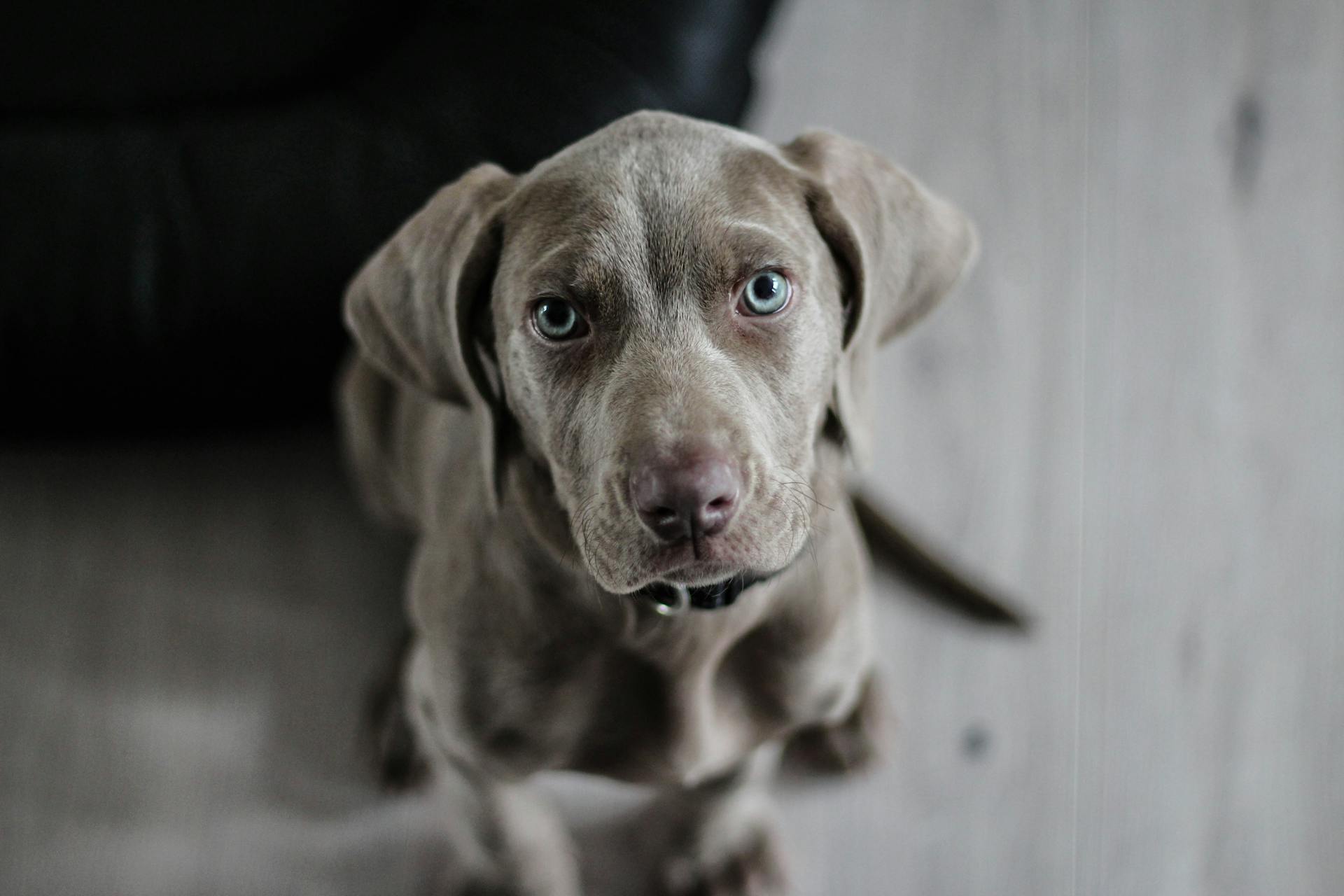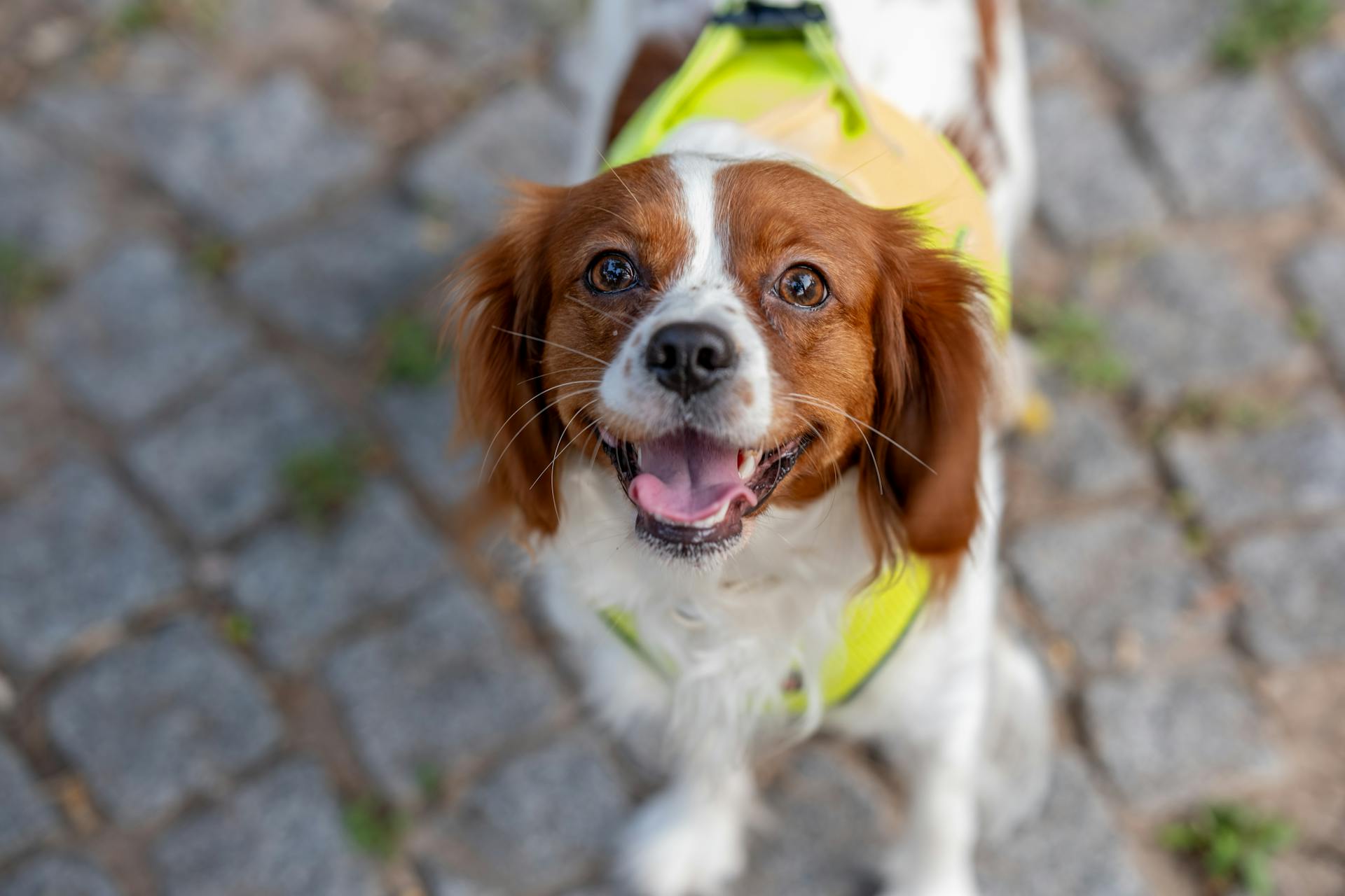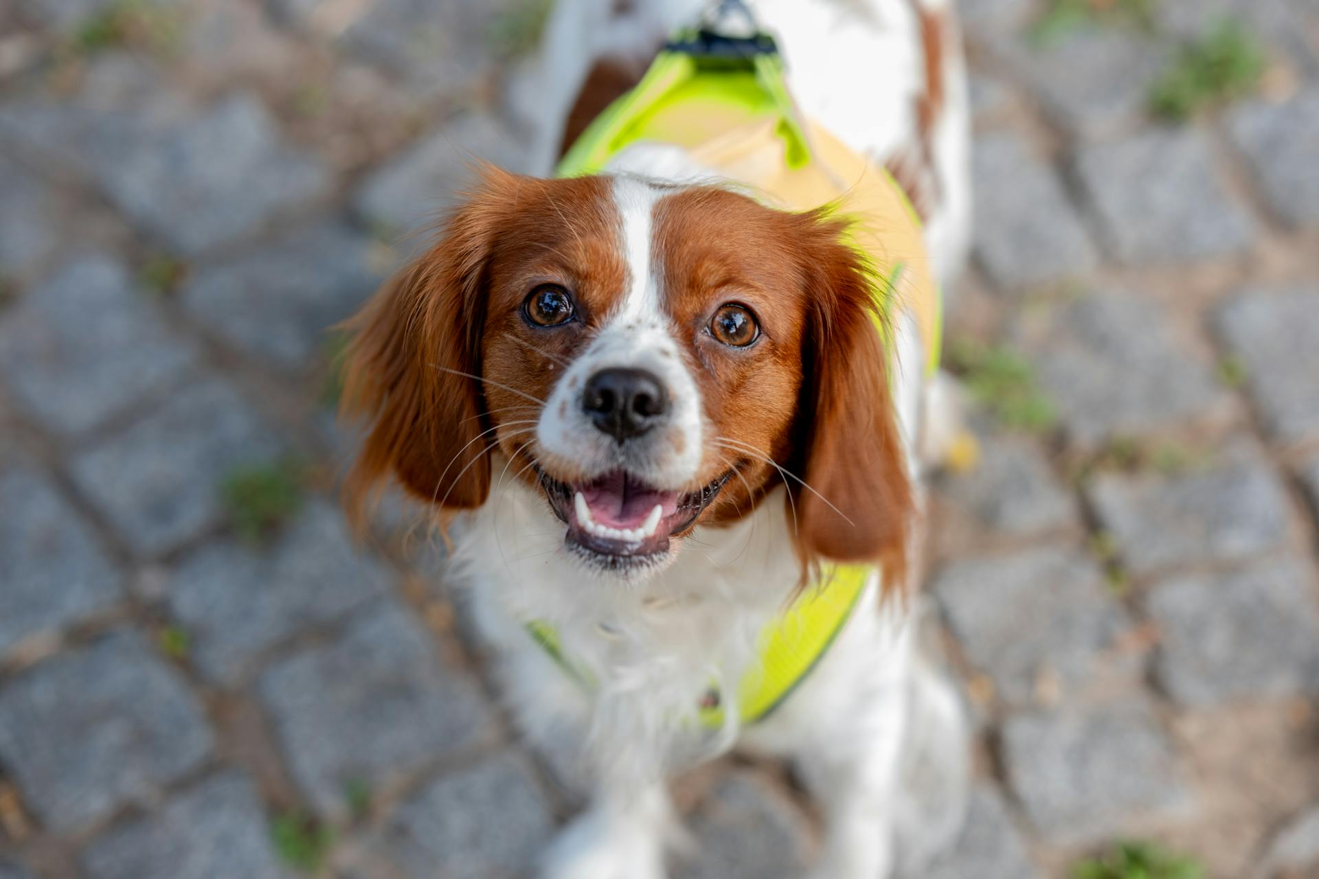
Let's start our journey from the top of our canine friend's head. The skull is made up of 22 bones that are fused together, and it's designed to protect the brain and sense organs. The eyes are positioned on either side of the head, and they're capable of moving independently of each other, allowing dogs to see almost 270 degrees around them.
The ears are triangular in shape and are extremely mobile, allowing dogs to pinpoint sounds with ease. They're also very sensitive to movement and can detect even the slightest changes in air pressure. Dogs use their ears to communicate and express emotions, and they can even fold them back against their head when they're feeling anxious or scared.
The nose is a highly sensitive organ that's responsible for detecting scents and pheromones. It's made up of millions of olfactory receptors that are capable of detecting even the faintest whiffs of smell. Dogs use their noses to gather information about their surroundings and to communicate with other dogs.
The mouth is equipped with 42 teeth, including incisors, canines, and molars. The teeth are designed for gnawing, tearing, and crushing food, and they're also used for defense and play-fighting.
Dog Body Parts
A dog's nose is often cold and wet, and it tends to get stuck in unwanted places.
The muzzle, comprised of the upper and lower jaws, is a distinctive feature of a dog's face. The stop, an indentation between the muzzle and forehead, can be nonexistent in some breeds.
The forehead, or braincase, is the portion of the head that resembles a human forehead, going from the stop and eyebrows to the back point of the skull. This area is a vital part of a dog's anatomy.
The occiput, the highest point of the skull at the back of the head, is a prominent feature on some dogs.
Here are the basic parts of a dog's face:
- Nose
- Muzzle
- Stop
- Forehead (braincase)
- Occiput
- Eyes
- Ears
- Brows
- Whiskers
- Flews (lips)
- Check
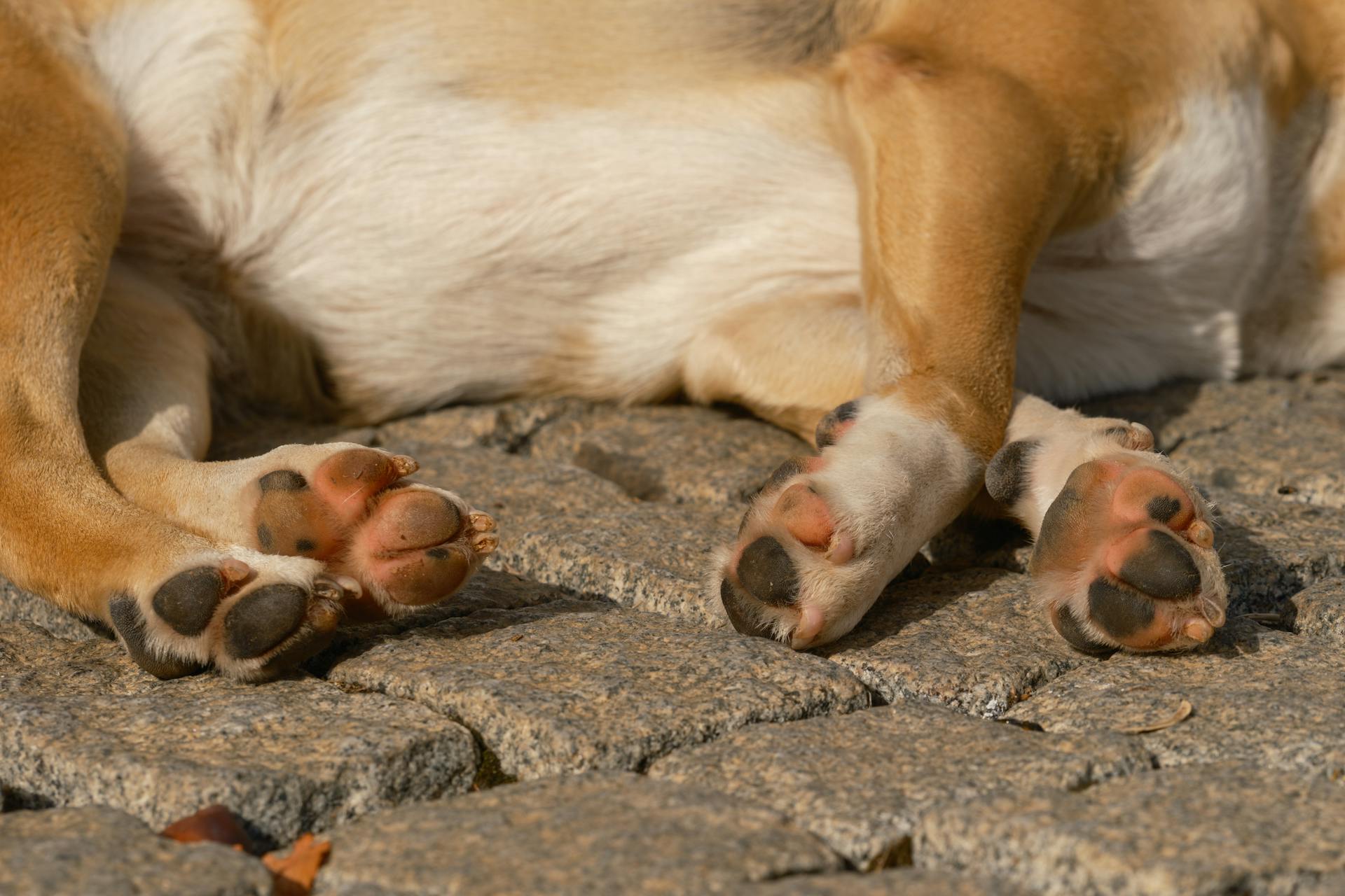
A dog's paw has five basic parts: the claw, digital pads, metacarpal (on the front paws) and metatarsal (on the rear paws) pad, dew claw, and carpal pad.
Check this out: Canine Paw Pad Anatomy
Forelegs
The forelegs of a dog are made up of several distinct parts. The upper arm, or humerus bone, is the long bone that runs from the shoulder to the elbow.
The elbow is the first joint in the foreleg, located just below the chest on the back of the foreleg. It's a crucial joint that allows the dog to bend and straighten its front leg.
The forearm, which is comprised of the ulna and radius bones, runs from the elbow to the wrist. You might notice feathering on the back of the forearm in some breeds.
The wrist, or lower joint, is located below the elbow on the foreleg. Below the wrist, you'll find the pasterns, which are equivalent to the bones in your hands and feet.
A dog's forefoot, or paw, is at the end of each foreleg and comes with nails (or claws), paw pads, and usually dewclaws. Dewclaws are vestiges of thumbs that dogs never developed.
Here's a breakdown of the parts of the forefoot:
- Main pad (communal pad)
- Pad under each toe
- Stopper pads behind the wrist
Each toe has a toenail or claw incorporated with part of the last bone of the toe.
Shoulder
The shoulder is a crucial part of the foreleg, and understanding its anatomy is essential for any dog owner. The shoulder is the top section of the foreleg, running from the withers to the elbow.
The withers are the top point of the shoulders, making them the highest point along the dog's back. This is a key landmark for measuring a dog's height and overall structure.
To better visualize the shoulder's location, consider the following breakdown: the shoulder starts at the withers and ends at the elbow. This is a critical area to examine when assessing a dog's overall health and mobility.
The shoulder's position and structure can greatly impact a dog's ability to move and perform daily activities. As an example, a dog with a well-developed shoulder is more likely to be agile and confident in its movements.
For more insights, see: Canine Elbow Anatomy
Forelegs
The foreleg is a fascinating part of a dog's anatomy. The upper arm on the foreleg is right below the shoulder and is comprised of the humerus bone, which is similar to the one found in your own upper arm. It ends at the elbow.
The elbow is the first joint in the dog's leg, located just below the chest on the back of the foreleg. It's a crucial joint that allows dogs to move their forelegs.
The long bone that runs after the elbow on the foreleg is the forearm. Like your arms, it's comprised of the ulna and radius. The forearm may have feathering on the back.
The wrist is the lower joint below the elbow on the foreleg. Sometimes called the carpals, pasterns are equivalent to the bones in your hands and feet — not counting fingers and toes — and dogs have them in both forelegs and hind legs.
Here's a breakdown of the foreleg bones:
- Humerus bone: the upper arm bone
- Elbow joint: the first joint in the dog's leg
- Forearm: the long bone that runs after the elbow, comprised of the ulna and radius
- Wrist (carpal): the lower joint below the elbow
- Pasterns: equivalent to the bones in your hands and feet
A dog's forefoot or paw is at the end of each foreleg, and it comes with nails, paw pads, and usually dewclaws.
A different take: Dogs Paw Anatomy
Hind Legs
The hind legs of a dog are a crucial part of their overall anatomy, allowing them to move, jump, and support their body weight.
A typical dog has two hind legs, each consisting of a thigh, a knee, a calf, and a paw.
Each hind leg is designed to work in tandem with the other, enabling the dog to move efficiently and effectively.
The hind legs are also responsible for supporting the dog's body weight, particularly when they're standing or sitting.
Expand your knowledge: Dog Coat Pattern Types
Pelvis and Hips
The pelvis plays a crucial role in locomotion and posture, protecting the pelvic viscera, reproductive, and urinary organs.
The pelvis is formed by two hip bones (ossa coxarum) that are joined ventrally at the cartilagenous pelvic symphysis and articulate dorsally with the sacrum.
Each hip bone is composed of three components: the ilium, pubis, and ischium. The ilium is the most cranial aspect of the hip, articulating with the sacrum.
The iliac crest is the outer margin of the ilium, representing an area of thickening around the rim of the ilium. It's a prominent feature that can be felt in many animals.
The gluteus medius muscles originate at the lateral surface of the ilium, helping to facilitate movement in the hind legs. I've seen this in action when watching dogs run and play.
The sciatic notch is a small area cut away from the wing of the ilium, allowing the sciatic nerve to pass over the pelvis to the hindlimb. This is a vital passage for nerve function.
The acetabulum is formed by all three bones and is surrounded by an acetabular rim. Its depth varies depending on breed, shape of pelvis, and hip conformation.
The pubis contributes to the formation of the cranial aspect of the acetabulum and the obturator foramen, through which the obturator nerve emerges. This is an important pathway for nerve function.
The ischium forms the caudal aspect of the pelvis and contributes to the acetabulum and the ischial tuber, a prominent horizontal thickening in many animals.
Upper Hindleg
The upper hindleg is a fascinating part of a dog's anatomy. The upper thigh is the part of the dog's leg situated above the knee on the hind leg.
Check this out: Bernese Mountain Dog Coat
The stifle or knee is the joint that sits on the front of the hind leg in line with the abdomen. This joint is a crucial part of the hind leg's structure.
The upper thigh is a key component of the hind leg, and it's worth noting that some dogs have feathering along the back of their upper thighs.
See what others are reading: Dogs Front Leg Anatomy
Paws and Bones
Dogs' paws work hard, and sometimes the elements can wear them down. This wear can be abrasion after long walks or runs on rough surfaces, leaving their normally rough and tough paw pads worn down and smooth.
Starting with short hikes and walks on rough terrain and slowly increasing that duration helps their paw pads develop tougher, calloused skin that's less prone to abrasion.
Dogs are digitigrade animals, meaning their weightbearing surface is their digits. The canine phalanges are crucial for their mobility.
The metacarpals in a dog's forelimb are numbered medially to laterally 1 to 5, corresponding to the numbers of the distal carpals.
Paw Health
A dog's paws work hard, and sometimes the elements can wear them down.
Walking or running on rough surfaces can cause abrasion, leaving paw pads worn down and smooth.
Short hikes and walks on rough terrain can help their paw pads develop tougher skin.
This is because their paw pads need to adapt to the new terrain, and with time, they'll become less prone to abrasion.
Tender paw pads can be a sign that your dog needs to take it easy on the hard or rocky terrain.
Starting with short walks and gradually increasing the duration can help prevent sore paw pads.
Regular walks on rough terrain can eventually lead to calloused skin that's less sensitive to rough surfaces.
You might enjoy: What Food Is Good for Dogs Skin and Coat
Forepaw Bones
Dogs are digitigrade animals, which means the weightbearing surface of their limbs is their digit. This makes their phalanges very important.
The canine phalanges are virtually identical in structure in the hindlimb and forelimb. The main differences are in the forelimb, where we have metacarpals and the metacarpophalangeal joint.
Worth a look: Canine Forelimb Anatomy
The metacarpals are numbered medially to laterally 1 to 5 in direct correspondence with the numbers of the distal carpals. Each metacarpal has a unique shape defined by its relative articulations.
The third and fourth carpal bones have a more square cross-section, as these middle metacarpals bear the majority of the weight borne by that limb. This is in contrast to metacarpals 2 and 5, which bear relatively less weight and have a more tringular cross-section.
Each metacarpal has a proximal region referred to as the base, a shaft or body, and a distal extremity or caput. The surface of the base of the metacarpal is relatively flat to provide articulation with the carpal bones.
Hindpaw Bones
Hindpaw bones are a crucial part of a cat's anatomy. They're made up of five toes, each with a specific bone structure.
The first toe, also known as the hallux, is a small, compact bone that's essential for balance and stability. It's also a key indicator of a cat's age, with younger cats having a more flexible hallux than older cats.
The hindpaw bones are connected to the leg bones by powerful muscles and tendons, allowing for smooth and agile movement. This is especially important for cats, as they need to be able to pounce and jump with ease.
Each toe has a unique bone structure, with the second toe being the longest and most flexible. This flexibility is essential for grasping and holding onto prey.
In cats, the hindpaw bones are also connected to the spine, allowing for a wide range of motion and flexibility. This is why cats are able to twist and turn with such ease.
Skull and Vertebrae
The canine skull and vertebrae are fascinating structures that play a crucial role in the dog's overall anatomy. The skull of a dog is a single bone, the cranium, which houses the brain and protects it from injury.
The canine vertebrae are divided into several regions, with the sacral vertebrae being a notable feature. The sacral vertebrae are found caudal to the lumbar vertebrae and are fused together to form a single bone, the sacrum. This fusion allows the dog to transmit locomotive force from the hindlimbs to the trunk of the body.
The sacrum is a key component of the canine vertebrae, narrowing caudally and curving to present a concave surface to the pelvic cavity.
Head and Neck
The head and neck area of a dog is quite fascinating. The nape of the neck is where the neck joins the base of the skull in the back of the head.
The throat is located beneath the jaws. This is an important distinction, especially when considering how to properly care for your dog's throat.
The crest starts at the nape and ends at the withers. The withers are the top point of the shoulders, making them the highest point along the dog's back.
The neck is pretty self-explanatory; it runs from the head to the shoulders. This is a crucial area to consider when evaluating a dog's overall health and structure.
Here's a quick rundown of the key terms:
- The nape of the neck: where the neck joins the base of the skull in the back of the head.
- The throat: located beneath the jaws.
- The crest: starts at the nape and ends at the withers.
- The neck: runs from the head to the shoulders.
- The withers: the top point of the shoulders.
Understanding these terms can help you better communicate with your veterinarian and make informed decisions about your dog's care.
Skull and Vertebrae
The sternum is a vital part of a dog's anatomy, and understanding its structure can be quite fascinating. It's a bony structure that runs along the thoracic cavity, providing attachment for the ribs and anchoring some of the muscles involved in respiration.
The sternum is composed of three elements: the manubrium, the body, and the xiphoid cartilage. The manubrium is rod-shaped and projects cranially to the first rib, and it can often be palpated at the region of the ventral neck.
The body of the sternum is generally cylindrical in shape and is composed of numerous segments interconnected by the costochondral ventral aspects of the ribs. These segments are referred to as sternebrae and are cartilaginous in young canines and ossify with age.
The xiphoid cartilage extends caudally in a similar manner to that of the manubrium cranially, providing attachments for the most cranial aspects of the abdomen and for the linea alba.
Here's a breakdown of the sternum's elements:
- Manubrium: rod-shaped, projects cranially to the first rib
- Body: cylindrical in shape, composed of numerous segments (sternebrae)
- Xiphoid cartilage: extends caudally, provides attachments for the abdomen and linea alba
Occipital Bone
The occipital bone is the most caudal bone of the skull.
It's a crucial part of the skull, providing the attachment for the nuchal ligament via the external occipital protuberance.
The occipital bone has nuchal crests laterally and a sagittal crest medially and dorsally to the external occipital protuberance.
The sagittal crest is prominent and can be palpated in most canines, unless they are very well muscled.
The nuchal wall, formed by these elements, also includes the foramen magnum.
The pars basilaris element is the caudal base of the cranium, with a cartilagenous suture joining it to the basisphenoid bone.
The muscular tubercules on the ventral surface of the pars basilaris provide attachment sites for the flexors of the head and neck.
The squamous part of the occipital bone is dorsal to the lateral parts and occipital condyles.
The nuchal crest is often used as a landmark for collecting cerebrospinal fluid.
The occipital condyles articulate with the atlas to form the atlanto-occipital joint.
The paracondylar process provides muscle attachment sites for the muscles of the head.
The hypoglossal canal is also located within the occipital bone.
Sacral Vertebrae
The sacral vertebrae in dogs are found below the lumbar vertebrae and consist of three fused vertebrae.
These vertebrae are fused together to form a single bone called the sacrum, which is a key part of the pelvic girdle.
The sacrum allows the locomotive force generated by the hindlimbs to be transmitted to the trunk of the dog.
In dogs, the sacrum narrows towards the back and is curved to fit snugly into the pelvic cavity.
A notable feature of the canine sacrum is that it retains small spinous processes, unlike some other species such as pigs.
In dogs, the three sacral vertebrae are usually fully fused to form a quadrilateral block.
The joint between the sacrum and the pelvis is typically made up of one or two sacral vertebrae in dogs.
Sources
- https://www.dummies.com/article/home-auto-hobbies/pets/dogs/health-grooming/dog-anatomy-from-head-to-tail-197577/
- https://canineanatomyforbeginners.wordpress.com/exterior-structure/
- https://ruffwear.com/blogs/explored/dog-paw-anatomy
- https://en.wikivet.net/Bones_-_Dog_Anatomy
- https://www.ukcdogs.com/canine-glossary
Featured Images: pexels.com


