Canine lymphoma cytology is a crucial diagnostic tool for veterinarians to accurately diagnose and treat canine lymphoma.
Cytology involves examining cells from a fine-needle aspirate or a tissue sample to identify abnormal cells.
In canine lymphoma cytology, abnormal lymphocytes are often identified, which can help diagnose the disease.
The morphology of these abnormal cells can vary, but they often appear as large, pleomorphic cells with irregular nuclei.
Accurate diagnosis is key to effective treatment, and cytology provides a rapid and minimally invasive way to obtain a diagnosis.
By understanding canine lymphoma cytology, pet owners and veterinarians can work together to develop an effective treatment plan.
Cytological Interpretation
Cytological Interpretation is a crucial step in diagnosing canine lymphoma.
If you're aspirating a lymph node, it's essential to follow guidelines to ensure accurate results.
There's generally a good correlation between cytologic and histologic diagnosis if the guidelines are adhered to.
Any enlarged lymph node may be aspirated to classify the lesion into one of three categories.
A normal lymph node is a typical result, indicating no lymphoma or other issues.
Reactive lymph nodes, also known as lymphoid hyperplasia, can occur due to infection, inflammation, or other stimuli.
Lymphoid neoplasia, or lymphoma, is a serious condition that affects the lymph nodes, often requiring prompt treatment.
Canine Lymphoma Diagnosis
Lymph node sampling is a quick and easy way to diagnose canine lymphoma. Fine-needle aspiration biopsy is a common method used to collect cytologic samples from lymph nodes.
To get an accurate diagnosis, it's essential to sample multiple lymph nodes, especially if they're enlarged. Sampling a lymph node away from the mouth or any site of inflammation can provide valuable information.
If the lymph nodes are not enlarged, lymph node cytology may not be helpful, as it's difficult to collect a sufficient sample. However, sampling a normal-sized lymph node can be done to investigate the potential for metastatic disease.
Cytological interpretation of lymph node aspirates can classify the lesion into one of three categories: normal lymph node, reactive lymphoid hyperplasia, or lymphoid neoplasia (lymphoma).
Lymph Nodes
Lymph node sampling and cytology is a quick and easy way to evaluate lymphadenopathy, which is the enlargement of lymph nodes.
A cytologic sample of a lymph node can be collected by fine-needle aspiration biopsy or nonaspiration fine-needle biopsy techniques.
Sampling can also be performed by imprints or scrapings from lymph nodes that have been surgically removed or at necropsy.
If multiple lymph nodes are enlarged, more than one should be sampled.
A lymph node away from the mouth or any site of inflammation should be aspirated as well as any lymph node close to a site of inflammation.
Lymph node cytology is not helpful if no lymph nodes are enlarged.
Normal sized lymph nodes may be aspirated on occasion to investigate the potential for metastatic disease.
Any enlarged lymph node may be aspirated for the purpose of classifying the lesion into a normal lymph node, reactive lymphoid hyperplasia, or lymphoid neoplasia (lymphoma).
Canine Lymphoma
Canine lymphoma is a serious condition that affects many dogs. The predominant cell type in canine lymphoma is the immature lymphoblast.
Lymphoblasts are large cells with nuclei that vary in size from 2 to 5 times the size of erythrocytes. Their cytoplasm is deeply basophilic and more abundant than that of small or intermediate lymphocytes.
The chromatin pattern of lymphoblasts is more diffuse and paler staining than in well-differentiated lymphocytes. A variable number of distinct or indistinct nucleoli are frequently visible.
The administration of glucocorticoids can drastically alter the lymphocytes population within a lymph node, making a lymphoma diagnosis difficult. This is because lymphoblasts are very sensitive to the cytotoxic effects of glucocorticoids.
Lymph node sampling and cytology is a quick and easy way to evaluate a lymphadenopathy. It can be performed by fine-needle aspiration biopsy or nonaspiration fine-needle biopsy techniques.
Lymph node cytology is an excellent way to evaluate a lymphadenopathy, whether it's a single, multiple, or generalized lymph node enlargement. It's essential to sample multiple lymph nodes if they're enlarged.
A lymph node away from the mouth or any site of inflammation should be aspirated, as well as any lymph node close to a site of inflammation. This helps ensure an accurate diagnosis.
If no lymph nodes are enlarged, lymph node cytology is generally not helpful. In such cases, aspiration of a nonenlarged node can be difficult and usually results in aspiration of perinodal fat with little or no lymphoid tissue present.
The most common physical examination finding in canine lymphoma is generalized peripheral lymphadenomegaly. This is seen in approximately 80% of patients with multicentric lymphoma.
Diagnostic Methods
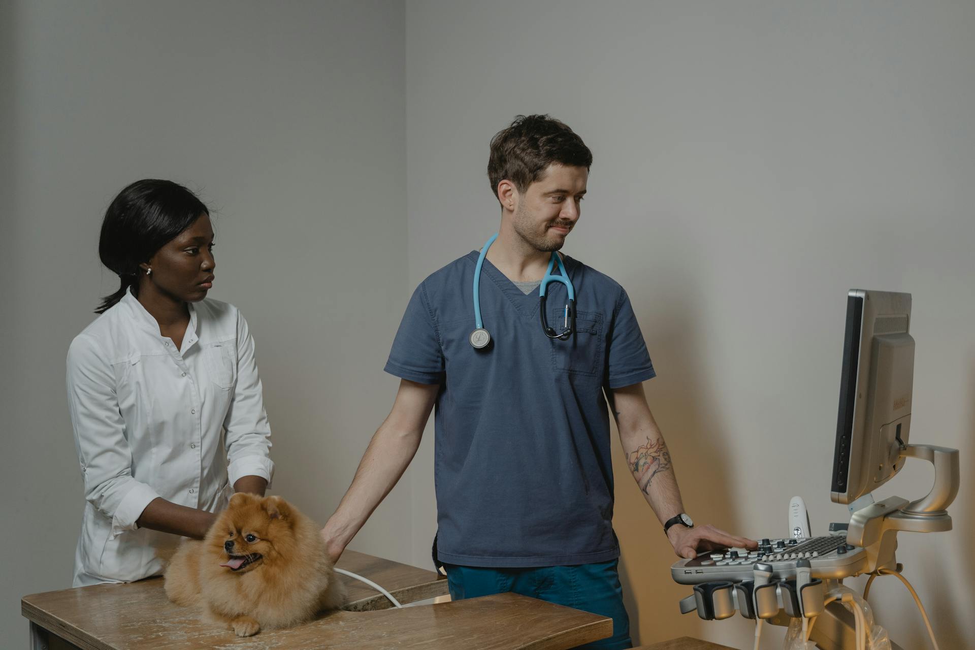
Fine-needle aspiration (FNA) is a minimally invasive and less expensive diagnostic method for canine lymphoma, providing rapid results in 1-2 days.
Cytology is typically performed on an affected lymph node or organ, and slides can be examined immediately in-house before being sent to a laboratory for confirmation by a pathologist.
The popliteal lymph nodes are easily accessible and a good starting point for FNA cytology, as they rarely require sedation.
Lymph node aspirates from the mandibular lymph nodes should be avoided due to their reactivity, as they drain the oral cavity.
Large cell (lymphoblastic) lymphoma is usually easily diagnosed via FNA cytology, but histopathology may be helpful in identifying and classifying indolent lymphoma, which is typically intermediate to small cell.
The whole lymph node should be excised and submitted for histopathology when cytology is inconclusive.
Sources
- https://www.dvm360.com/view/cytology-evaluation-lymphoid-tissue-dog-and-cat-proceedings
- https://pubmed.ncbi.nlm.nih.gov/27487521/
- https://air.unimi.it/handle/2434/874015
- https://www.cliniciansbrief.com/article/canine-multicentric-lymphoma
- https://www.ksvdl.org/resources/news/diagnostic_insights/may2020/diagnosing-canine-lymphoma.html
Featured Images: pexels.com