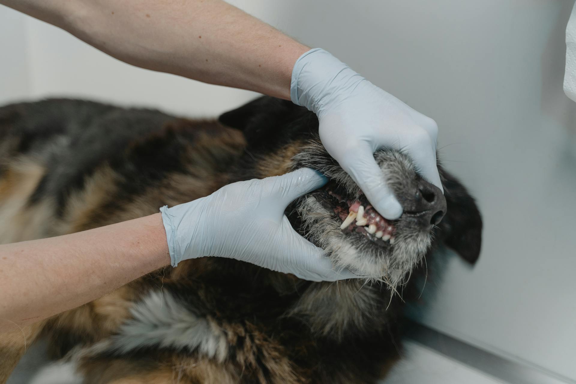
Canine oral cancer can be a sneaky and aggressive disease, making it essential to know the signs and symptoms to catch it early.
One of the most common signs of oral cancer in dogs is a mass or lump in the mouth, which can be red, white, or black in color.
Oral cancer can also cause swelling in the jaw or face, difficulty eating or swallowing, and bad breath.
The most common type of oral cancer in dogs is squamous cell carcinoma, which can appear as a white patch or ulcer in the mouth.
Symptoms can vary depending on the location and size of the tumor, but owners should be aware of any changes in their dog's behavior or physical appearance.
Early detection is key, and regular veterinary check-ups can help identify potential issues before they become serious problems.
Take a look at this: Food for Anemic Dogs
Common Symptoms
Pets with oral tumors may have a history of pain while trying to chew or swallow food. This pain can be severe enough to make eating a chore.
Additional reading: Canine Cancer Pain Management
Food may drop out of the mouth while eating, or pets may not be willing to eat at all. I've seen dogs with oral tumors avoid their favorite treats.
Drooling is another common symptom, as pets try to compensate for the discomfort in their mouth. Some dogs may even have blood-tinged saliva if the tumor is ulcerated.
Periodontal disease, bad breath, and tooth loss may also be present. These symptoms can be a sign of an underlying oral tumor.
By the time owners notice any abnormality, the mass is likely to be large enough to be visualized in the oral cavity. Regular dental check-ups can help catch these tumors early.
Curious to learn more? Check out: Canine Mouth Cancer Life Expectancy
Diagnosis and Histology
A board-certified pathologist reviewed hematoxylin and eosin slides to confirm the diagnosis and ensure the presence of viable tumor tissue.
Immunohistochemistry was performed using a Leica Bond Max autostainer with a panel of antibodies against CD3, CD204, CD79a, and FoxP3.
The antibodies were used to identify pan T lymphocytes, macrophages, B lymphocytes, and regulatory T cells, respectively.
Antigen retrieval was performed using either Tris-EDTA buffer or Citrate buffer, depending on the antibody being used.
Whole slide brightfield images of IHC stained slides were digitally captured at 10x magnification and used for quantitative analysis of tumor-infiltrating leukocyte density.
Common Oral Tumors in Dogs
Oral melanoma in dogs can be treated with maxillectomy and chemotherapy, resulting in a median survival time of 4-12 months.
This type of tumor is highly dependent on clinical stage of disease, which significantly affects prognosis.
Surgery alone or radiation therapy alone typically yields a median survival time of 4-12 months for dogs with oral melanoma.
However, combining surgery with either chemotherapy or immunotherapy may lead to a longer survival time.
Oral fibrosarcoma in cats can be fatal, often causing the cat to be euthanized due to the size of the mass.
Recommended read: Images of Toy Dogs
Oral squamous cell carcinoma in dogs has a median survival time of 9-26 months with surgery alone.
Adding radiation therapy or chemotherapy does not seem to improve prognosis for this type of tumor.
Canine oral squamous cell carcinoma can be treated with maxillectomy, and in some cases, the dog may live for 18 months or more before dying of unrelated causes.
Oral fibrosarcoma in dogs has a reported median survival time of 7-12 months with surgery alone.
Local recurrence is reported in up to 57% of dogs with this type of tumor.
However, when treated with a combination of surgery and radiation therapy, the median survival time for dogs with oral fibrosarcoma increases to 18-26 months.
A unique perspective: Canine Oral Anatomy
Histology and IHC
Histology and IHC are crucial steps in confirming a diagnosis and understanding the presence of viable tumor tissue.
A board-certified pathologist reviewed hematoxylin and eosin slides to confirm diagnosis and the presence of adequate viable tumor tissue prior to immunohistochemical labeling.
Immunohistochemistry was performed using a Leica Bond Max autostainer with a panel of previously published canine cross-reactive primary antibodies directed against specific antigens/cell types.
The panel included monoclonal mouse anti-human CD3, monoclonal mouse anti-human CD204, mouse monoclonal anti-human CD79a, and mouse monoclonal anti-human FoxP3.
Antigen retrieval was performed using either Leica Epitope Retrieval 2 or Leica Epitope Retrieval 1, depending on the antibody used.
Detection was performed with PowerVision IHC detection systems, using a polymeric horseradish peroxidase anti-mouse IgG and Bond Polymer Refine DAB chromogen, with routine hematoxylin counterstain.
Quantitative analysis of tumor-infiltrating leukocyte density was performed using whole slide brightfield images of IHC stained slides digitally captured using an Olympus IX83 microscope.
Images were converted to gray scale images for analysis, and tumor tissue regions-of-interest were segmented from adjacent normal tissue by manual annotation in ImageJ.
Positively labeled immune cells were quantified using the color deconvolution algorithm, with a positive pixel threshold determined visually by a veterinary pathologist.
A fresh viewpoint: Images of Boykin Spaniels
Fig. 4
Fig. 4 is a crucial image in understanding canine oral cancer, and it shows a tumor growing on the roof of a dog's mouth.
This tumor is a squamous cell carcinoma, which is the most common type of oral cancer in dogs.
The image highlights the importance of early detection, as the tumor is still relatively small and could be treated more effectively if caught sooner.
The dog in the image is a 10-year-old golden retriever, and its owner noticed a foul odor coming from its mouth, which led to a veterinary visit.
Squamous cell carcinoma often presents with symptoms like bad breath, difficulty eating, and swelling in the face or neck.
The image is a great example of how oral cancer can affect a dog's quality of life, and why regular check-ups with a veterinarian are essential.
Frequently Asked Questions
What is the life expectancy of a dog with oral cancer?
The life expectancy of a dog with oral cancer varies from 7-40 months, depending on the treatment approach. With surgery and radiation, dogs can live up to 10-40 months, while chemotherapy alone averages 7-8 months.
What is the most aggressive oral cancer in dogs?
Tonsillar SCC is the most aggressive oral cancer in dogs, with a high risk of metastasis to local lymph nodes and other parts of the body
Sources
- http://www.veterinarycancer.com/oral-tumors
- https://www.ncbi.nlm.nih.gov/pmc/articles/PMC8189657/
- https://www.frontiersin.org/journals/oncology/articles/10.3389/fonc.2022.1033704/full
- https://hospital.cvm.ncsu.edu/services/small-animals/cancer-oncology/oncology/canine-oral-melanoma/
- https://www.cliniciansbrief.com/article/common-oral-tumors-dogs-cats
Featured Images: pexels.com