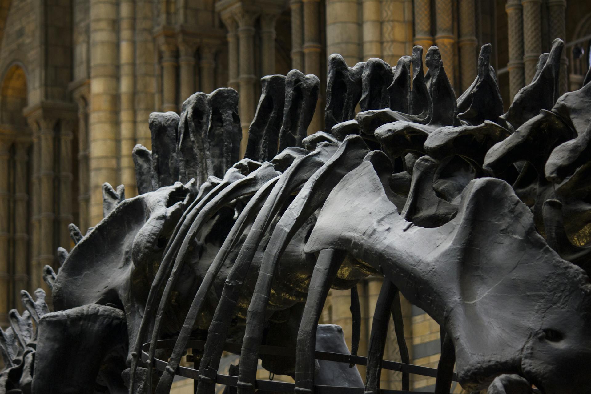
Understanding the canine pelvis anatomy is crucial for any dog owner or enthusiast. The canine pelvis is a complex structure composed of three bones: the ilium, ischium, and pubis.
The ilium is the largest and widest of the three bones, forming the majority of the pelvis. It has a distinctive wing-like shape, which provides a wide base for the pelvis.
The canine pelvis plays a vital role in supporting the body's weight and facilitating movement. It also serves as a crucial attachment point for muscles, tendons, and ligaments.
Pelvic Anatomy
The canine pelvis is a complex structure that plays a crucial role in supporting the body's weight and facilitating movement. It's made up of several key components, including the pelvic symphysis, sacroiliac joints, and sacrotuberous ligament.
The pelvic symphysis is a secondary cartilaginous joint that ossifies with age and may expand in parturition. This means that as a dog gets older, the symphysis will gradually turn into bone, and during pregnancy, it may widen to accommodate the growing fetus.
The sacroiliac joints are a unique combination of synovial and fibrous joints, which allows them to transmit the weight of the trunk to the hindlimbs. This is essential for supporting the dog's body weight and enabling movement.
Here are the key components of the canine pelvis:
Understanding the anatomy of the canine pelvis is essential for veterinarians and dog owners to diagnose and treat various conditions that affect the pelvis and surrounding areas.
Pelvic Joints and Ligaments Core
The pelvis is a complex structure composed of several joints and ligaments that work together to support the body's weight and facilitate movement. The pelvic symphysis is a secondary cartilaginous joint that ossifies with age and may expand in parturition.
The sacroiliac joints are a unique combination of synovial and fibrous joints, playing a crucial role in transmitting the weight of the trunk to the hindlimbs. This joint is essential for the overall stability and mobility of the pelvis.
The sacrotuberous ligament is quite variable between species, and its caudal edge can be palpable. This ligament is likely to be more prominent in certain breeds or individuals, depending on their specific anatomy.
Here's a quick rundown of the key pelvic joints and ligaments:
Understanding the anatomy of the pelvis is essential for any veterinary professional or animal enthusiast. By grasping the intricacies of the pelvic joints and ligaments, we can better appreciate the remarkable complexity of the animal body.
Species Differences
One of the most fascinating aspects of pelvic anatomy is the way it differs between species. Larger species have a more vertical ilium, which brings the sacroiliac joint and the weight of the trunk closer to the hip.
This design allows them to support their own body weight more efficiently. The dog, for example, has a stout cord extending between the sacrum and lateral ischial tuberosity, which is not present in the cat.
In smaller species like cats, the ilium is more oblique, giving them a slightly different gait and balance. This is likely an adaptation to their smaller size and lighter body weight.
The ungulates, on the other hand, have a unique feature - their sacrosciatic ligament expands to a broad sheet. This allows them to distribute the weight of their body more evenly, which is essential for their distinctive gait.
You might enjoy: Dog Gait Types
Canine Anatomy on CT
Imaging studies like CT scans can provide a detailed look at the canine pelvis anatomy, including the ilium, ischium, and pubis bones. The ilium is the largest and widest of these bones, making up the majority of the pelvis.
The CT scan images show that the canine pelvis is designed for mobility and flexibility, with the acetabulum forming a deep socket to support the femoral head. The pelvis is also composed of a ring of bones that surround the acetabulum, providing additional support.
The CT scan results show that the canine pelvis has a unique structure that allows for a wide range of motion, making it an essential part of a dog's overall mobility and flexibility.
Expand your knowledge: Norwegian Lundehund Flexibility
Male Canine Abdomen Anatomy on CT
The male canine abdomen anatomy on CT is a fascinating topic. The stomach is a muscular sac that can hold up to 1 liter of food and water, as seen in CT scans.
The liver is a vital organ that plays a crucial role in detoxification and metabolism, weighing around 100-150 grams in a typical male dog. It's located in the upper right quadrant of the abdomen.
The pancreas is a gland that produces digestive enzymes and hormones, sitting behind the stomach and in front of the spleen. In a CT scan, it's visible as a thin, elongated structure.
The spleen is an essential organ that filters the blood and stores red blood cells, weighing around 50-100 grams in a typical male dog. It's located in the upper left quadrant of the abdomen, near the stomach.
The kidneys are two bean-shaped organs that filter waste and excess fluids from the blood, weighing around 50-70 grams each in a typical male dog. They're located on either side of the spine.
You might enjoy: Male Dogs Nipple
Results
Analyzing the results of our canine anatomy study on CT scans, we found that the Norberg angle didn't show significant associations when averaged over both hips.
The first principal component (PC1) was negatively correlated with the Norberg angle, with a correlation coefficient of -0.31 and a P-value less than 0.05.
PC1, 2, 4, and 5 showed significant differences between male and female dogs, confirming the presence of pelvic sexual dimorphism in canines.
The eigenvector contribution to PC1 was significantly associated with the overall size of the pelvis, and with the IGF-1 locus, a known contributor to canine body size.
PC3 represented a tradeoff between ilial length and ischial length, and was significantly associated with a marker on canine chromosome 16:5181388 bp.
The closest candidate gene associated with PC3 is TPK1, a thiamine-dependent enzyme and part of the PKA complex.
Featured Images: pexels.com