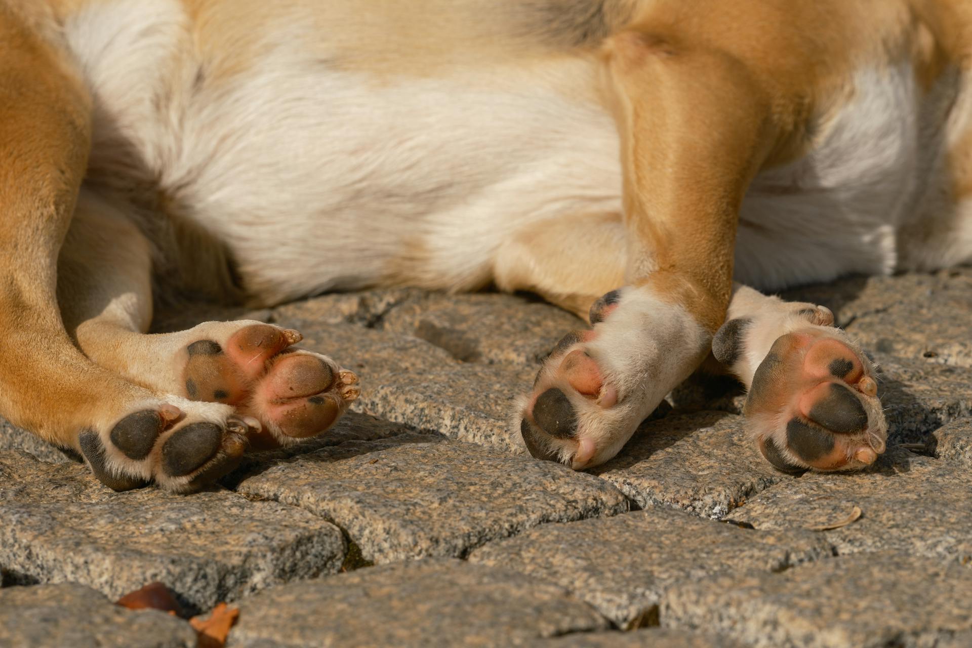
The canine tarsus is a complex structure that's essential for a dog's mobility and balance. It's composed of seven bones, including the calcaneus, talus, cuboid, and four tarsal bones.
The tarsal bones are arranged in two rows, with the proximal row consisting of the calcaneus and talus, and the distal row consisting of the cuboid and four tarsal bones. This arrangement allows for a wide range of motion in the hind legs.
The calcaneus, or heel bone, is the largest bone in the tarsus and plays a crucial role in weight-bearing and shock absorption. Its unique shape and size make it an essential component of the canine tarsus.
The talus, or ankle bone, is the second largest bone in the tarsus and is responsible for transmitting forces from the femur to the tarsal bones, allowing for smooth movement and weight-bearing.
Methods
To understand canine tarsal anatomy, it's essential to know the methods used to study and analyze the tarsal bones.
The most common method is radiography, which allows for clear visualization of the tarsal bones and their surrounding structures.
Radiography involves taking X-rays of the affected area, providing valuable information about the bone's shape, size, and any potential fractures or abnormalities.
This method is particularly useful for identifying issues such as osteochondritis dissecans, a condition characterized by the breakdown of cartilage and bone in the tarsal joints.
Ultrasonography
To perform ultrasonography, the hair is clipped from the midtibia to the level of the metatarsophalangeal joint.
The ultrasonography is done using a 12-MHz linear transducer. This specific type of transducer is used for its high frequency and linear shape, allowing for clear images of the tarsus.
The scans are performed in a specific sequence: dorsal, plantar, lateral, and medial. This order helps to ensure that all areas of the tarsus are thoroughly examined.
The dog is positioned in different recumbencies for each portion of the tarsus. This includes dorsal recumbency for the dorsal and plantar portions, right lateral recumbency for the right medial and left lateral portions, and left lateral recumbency for the left medial and medial portions.
During scanning, the probe is moved from the proximal to distal aspect and from the medial to lateral aspect. This helps to capture a comprehensive view of the tarsus.
Image Analysis
Image analysis was conducted by two veterinarians with 2 years of radiology experience, who assessed MRI images on a PACS System Elva.
The veterinarians used identical evaluation criteria and rated utility on a 5-point scale for each structure, considering degree of structural visualization and reliability.
MRI images were transferred and assessed simultaneously, and the evaluation order of the structures was random.
The evaluation was conducted in three planes: sagittal, dorsal, and transverse, using three sequences: T1W, T2W, and PD FS on MRI.
Ultrasonography was performed by one veterinarian and concurrently assessed by the same two observers who evaluated the MRI images.
The scanning sequence for ultrasonography included four approaches: dorsal, plantar, lateral, and medial, and was conducted in both transverse and longitudinal views.
Hock Anatomy
The hock is a complex joint that's prone to various issues. It's made up of several joints, including the tibiotarsal or tarsocrural joint, proximal intertarsal joint, distal intertarsal joint, tarsometatarsal joint, and talocalcaneal joint.
These joints are supported by a group of bones, including the talus, calcaneus, central tarsal bone, fused 1st and 2nd tarsal bone, 3rd tarsal bone, 4th tarsal bone, 2nd metatarsal bone, 3rd metatarsal bone, and 4th metatarsal bone.
A horse's hock can be affected by "capped hock", a condition caused by the creation of a false bursa beneath the skin, usually due to trauma. This is often of cosmetic significance and doesn't require immediate veterinary attention.
Osteochondrosis dissecans, or OCD, is a developmental defect in the cartilage or cartilage and bone seen in the tarsocrural joint. It's a common cause of bog spavin and can be treated with surgery.
Distension of the tibiotarsal joint with excessive joint fluid and/or synovium is called bog spavin. This can be a sign of OCD or other underlying issues.
Here are the joints of the hock:
- Tibiotarsal or tarsocrural joint
- Proximal intertarsal joint or talocalcanealcentroquartal joint
- Distal intertarsal joint or centrodistal joint
- Tarsometatarsal joint
- Talocalcaneal joint
And here are the bones of the hock:
- Talus
- Calcaneus
- Central tarsal bone
- Fused 1st and 2nd tarsal bone
- 3rd tarsal bone
- 4th tarsal bone
- 2nd metatarsal bone
- 3rd metatarsal bone
- 4th metatarsal bone
Featured Images: pexels.com

