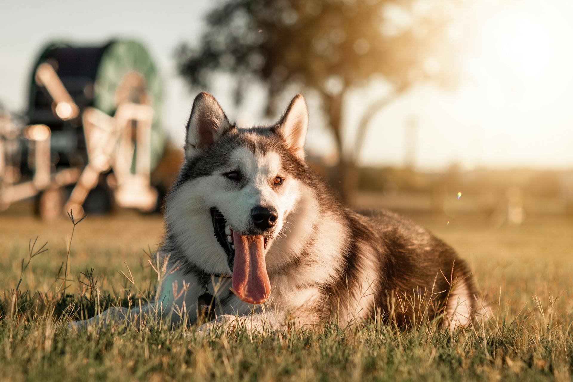
The canine tongue is a remarkable feature, and understanding its anatomy is key to appreciating its importance in a dog's daily life.
The canine tongue is made up of a muscular structure that is attached to the bottom of the mouth, with a unique shape that allows it to move in a wide range of motions.
This muscular structure is composed of two main parts: the intrinsic and extrinsic muscles. The intrinsic muscles are responsible for the tongue's movement and shape, while the extrinsic muscles control its position and attachment to the jaw and hyoid bone.
The tongue's surface is covered with small bumps called papillae, which help to collect and manipulate food particles.
Broaden your view: Dogs Tongue White
Canine Tongue Anatomy
A dog's tongue is made up of skeletal muscle, mucous membrane, glands, nerves, and vessels, giving it the unique texture and function we see in our furry friends.
The dog tongue has three main parts: the root, a long body, and apex. The root is attached to the hyoid bone and mandible by muscles.
Expand your knowledge: Why Do Chihuahuas Stick Their Tongue Out
The body of the dog tongue has a rough dorsal surface due to the presence of different types of papillae, such as filiform, fungiform, vallate, foliate, and conical.
At the ventral surface, you'll find an unpaired mucosal fold (lingual frenulum) connecting the body of the tongue to the floor of the mouth cavity.
Consider reading: Chowchow Tongue
Gross Anatomy
A dog's tongue is a fascinating part of their anatomy, and understanding its gross anatomy can be really helpful in appreciating their unique features.
The root of a dog's tongue is very thick and attached to the hyoid bone and mandible by hyoglossus and genioglossus muscle.
The root of a dog's tongue is also fixed to the ventral surface, while the dorsal surface is free. This is a key distinction in the anatomy of a dog's tongue.
The body of a dog's tongue is elongated and presents two surfaces: the dorsal surface, also known as the dorsum linguae, and the ventral surface.
The dorsal surface of a dog's tongue is rough due to the presence of different types of papillae, including filiform, fungiform, vallate, foliate, and conical papillae.
A median groove divides the dorsal surface of a dog's tongue into two lateral halves. This is a unique feature of a dog's tongue compared to other animals.
The ventral surface of a dog's tongue is smooth, except for an unpaired mucosal fold known as the lingual frenulum that connects the body of the tongue to the floor of the mouth cavity.
The tip of the ventral surface of a dog's tongue presents a thick fibromuscular cord-shaped structure known as the lyssa.
Canine
The canine tongue is a remarkable feature of our furry friends. It's heavily keratinized, which means it's tough and resistant to wear and tear.
This keratinization helps protect the tongue while eating, and it's especially evident in the long papillae that cover the tongue. These papillae are like tiny little hairs that help pick up food.
The ox has a similar type of papillae, called lenticular papillae, which are hard and horny due to heavy keratinization. This is a great example of how different animals have adapted their tongues to suit their needs.
Most of the papillae on a canine tongue are soft and long, and they point downwards, or caudally. This unique shape helps the tongue move food around efficiently.
Mouth
The mouth is a vital part of a dog's anatomy, playing a crucial role in their daily life. It's located in the front, lower part of the face and contains a tongue, teeth, and gums.
The mouth is surrounded by cheeks and lips, which help to keep food in place while eating. The entire mouth is lined with mucus membranes that manufacture and secrete saliva, keeping the mouth moist and helping to move debris and food down into the throat and digestive tract.
A dog's mouth also acts as an airway, in addition to the nose. This means that they will use their mouth to breathe more than their nasal passage when exercising, becoming excited, or if their nose is blocked.
The mouth is the entrance to the gastrointestinal tract, where food is broken down and absorbed. It's a remarkable system that allows dogs to eat and digest their food efficiently.
You might enjoy: Canine Food Bloat
Tongue Types
There are several types of tongues found in canines, each with its unique characteristics. The most common types are the flat tongue, the rolled tongue, and the fringed tongue.
A flat tongue is typically found in breeds such as the Greyhound and the Whippet, and is characterized by its broad, flat shape. This type of tongue is well-suited for these breeds' sighthound ancestry.
The rolled tongue, on the other hand, is found in breeds such as the Bulldog and the Pug, and is characterized by its rolled or curled shape. This type of tongue is often seen in breeds with brachycephalic (short-nosed) skulls.
Worth a look: Rear Dew Claws on Dogs Breeds
Conical
The conical papillae are a type of papillae found on the dog tongue. They are conical in shape and have a wide circular base that narrows to a thin apex.
Each conical papillae has a caudally directed apex that is heavily cornified than the base. The largest conical papillae are found just caudal to the palatoglossal arch.
The dorsal surface of the conical papillae is lined with stratified squamous epithelium. Some conical papillae are modified to form the wall of the outer moat of the vallate papillae.
Conical papillae possess mechanical and tactile functions rather than gustatory functions. They are innervated by the glossopharyngeal nerve.
Here are some key characteristics of conical papillae:
Filiform
Filiform papillae are the most numerous type of papillae on a dog's tongue, covering the rostral two-thirds of the tongue body.
They are the smallest papillae, with a complex structure consisting of a broad base and a dermal core.
The primary filiform papillae are single and located centrally, with a dermal core forming their axis.
Four to six secondary filiform papillae surround the base of the primary papillae, arising from them.
Tertiary filiform papillae arise from the secondary papillae, without a dermal core, and are smaller than the secondary ones.
They are made up of epithelial tissue and have a well-cornified surface to protect deeper structures.
Filiform papillae are also found in cats, where they are very prominent and present in the rostral 2/3 of the tongue.
They consist of a thick keratin on stratified squamous epithelium and do not have taste buds, glands, or lymphatics.
Foliate
The foliate papillae are a type of tongue feature found in dogs. They are located on the dorsolateral aspect of the caudal third of the tongue.
Each group of foliate papillae consists of eight to thirteen papillae, which are covered with stratified squamous epithelium. This type of epithelium is also known as cornified.
The long axis of each papilla runs obliquely from the side of the tongue towards the dorsum, in a radiating manner. This unique shape allows for efficient sensory reception.
Foliate papillae are bordered dorsally, rostrally, and caudally by conical papillae, but not in the lateral boundary of the group. This distinct arrangement suggests a specialized function.
A central primary dermal core is present in each individual foliate papilla, from which a secondary dermal papilla arises.
Marginal
Marginal papillae are a unique feature found in the tongues of newborn dogs.
They're distributed along the margin of the rostral half of the tongue, with well-developed papillae at the level of the premolar.
Less developed papillae are present at the margin of the apex of the dog tongue.
You won't find any marginal papillae at the margins of the caudal half of the tongue.
These marginal papillae are threadlike and narrower at their apices.
Their function is mechanical and tactile rather than gustatory, meaning they're not involved in taste.
Types of
Types of papillae on the dog tongue are numerous and complex, but they all play a vital role in the dog's sensory experience.
There are three main types of papillae: filiform, vallate, and foliate. Filiform papillae are the smallest and most numerous, found on the rostral two-thirds of the tongue. They consist of a broad base and a dermal core, with three different types: primary, secondary, and tertiary.
Vallate papillae are larger and fewer in number, found at the caudal third of the tongue. They are arranged in a V shape and can vary in number depending on the breed of dog. There are two types of vallate papillae: simple and complex.
Explore further: Dog Tail Types
Foliate papillae are found on the dorsolateral aspect of the caudal third of the tongue and are covered with stratified squamous epithelium. They are arranged in groups and have a central primary dermal core with a secondary dermal papilla.
Conical papillae are also found on the tongue and are located on the dorsum of the caudal third. They are conical in shape and have a wide circular base that narrows to a thin apex.
Each type of papillae has a unique structure and function, but they all contribute to the dog's ability to taste, feel, and experience its environment.
Tongue Muscles
The tongue muscles in a dog are responsible for various functions, including movement, protrusion, and depression. The extrinsic muscles, which are the most lateral, include the styloglossus, hyoglossus, and genioglossus muscles.
The styloglossus muscle is the most lateral of the three extrinsic muscles and has three distinct heads: a short head, a rostral head, and a long head. The short head arises from the distal half of the caudal surface of the hyoid bone. The long head of the styloglossus muscle arises from the stylohyoid bone immediately dorsolateral to the origin of the short head.
For another approach, see: Dogs Muscle Anatomy
The hyoglossus muscle is another lateral extrinsic lingual muscle that arises from the ventrolateral surface of the basihyoid and thyrohyoid bones. It runs dorsal to the mylohyoideus muscle and lateral to the genioglossus muscle. The hyoglossus muscle retracts and depresses the dog's tongue.
Here are the main extrinsic muscles of the dog tongue:
- Styloglossus muscle
- Hyoglossus muscle
- Genioglossus muscle
The genioglossus muscle is fan-shaped and has three distinct bundles: a vertical bundle, an oblique bundle, and a straight bundle. The vertical bundle arises from the ventromedial surface of the rostral end of the mandible. The oblique bundle lies caudal to the vertical bundle and originates from the ventromedial aspect of the mandible.
Styloglossus Muscle
The styloglossus muscle is the most lateral extrinsic muscle of the dog tongue anatomy, located at the caudal third of the tongue.
You'll find a distinctive narrow proximal end and wide rostral end in the styloglossus muscle of the tongue.
There are three different heads present in the styloglossus muscle of the dog tongue: the short head, the rostral head, and the long head.
Here are the specific origins of each head:
- The short head arises from the distal half of the caudal surface of the hyoid bone.
- The rostral head arises from the rostrodorsal surface of the proximal half of the stylohyoid bone.
- The long head arises from the stylohyoid bone immediately dorsolateral to the origin of the short head.
The styloglossus muscle fibers of the long head curve ventrally and rostrally along with the ventral surface of the tongue.
Hyloglossus Muscle
The hyloglossus muscle is another lateral extrinsic lingual muscle of a dog, located at the root of the tongue below the styloglossus muscle.
It arises from the ventrolateral surface of the basihyoid and thyrohyoid bones.
This muscle runs dorsal to the mylohyoideus muscle and lateral to the genioglossus muscle.
The hyloglossus muscle crosses the medial aspect of the styloglossus muscle at the root of the tongue.
This muscle retracts and depresses the dog's tongue.
The hyloglossus muscle is innervated by the hypoglossal nerve.
Genioglossus Muscle
The genioglossus muscle is a fan-shaped muscle located on the dog's tongue.
This muscle is situated dorsal to the geniohyoideus muscle and has two distinct ends - a narrower end and an expanded end.
The narrower end of the genioglossus muscle originates from the medial surface of the mandible adjacent to the intermandibular joint.
The expanded end of the muscle inserts into the ventral surface of the dog's tongue.
The genioglossus muscle is composed of three distinct bundles of muscle fibers: verticle, oblique, and straight.
The verticle bundle of muscle fibers is located at the rostral part of the genioglossus muscle and inserts on the rostral half of the dog's tongue.
This verticle bundle arises from the ventromedial surface of the rostral end of the mandible.
The oblique bundle of muscle fibers lies caudal to the verticle bundle and inserts on the caudal half of the tongue.
The straight bundle of the genioglossus muscle lies lateral to both the verticle and oblique bundles.
The genioglossus muscle helps to depress and protrude the tongue.
It is innervated by the hypoglossal nerve.
Extrinsic Muscles
The extrinsic muscles of a dog's tongue play a crucial role in its movement and function. They are located on the outside of the tongue and are responsible for its protrusion and local movements.
The proper lingual extrinsic muscle forms the core of the extrinsic group and has four types of fibers. These fibers are categorized into superficial longitudinal, deep longitudinal, transverse, and perpendicular fibers.
The superficial longitudinal fibers are well-developed at the caudal half of the tongue, located immediately deep into the dorsal lingual mucosa. They help in protruding the tongue.
The deep longitudinal fibers, on the other hand, are less numerous and organized, located in the ventral half of the tongue. They are found in compact masses at the rostral end of the tongue and surround the lyssa body.
Here's a breakdown of the types of fibers found in the proper lingual extrinsic muscle:
- A superficial longitudinal fiber of proper lingual muscle
- The deep longitudinal fiber of the proper lingual muscle
- A transverse fiber of proper lingual muscle
- The perpendicular fibers of the proper lingual muscle of the dog tongue
These extrinsic muscles not only help in the movement of the tongue but also prevent it from being bitten.
Sources
- https://anatomylearner.com/dog-tongue-anatomy/
- https://en.wikivet.net/Tongue_-_Anatomy_%26_Physiology
- https://www.petplace.com/article/dogs/pet-health/structure-and-function-of-the-tongue-teeth-and-mouth-in-dogs
- https://thepetlabco.com/learn/dog/health-wellness/dogs-mouth
- https://www.tumblr.com/puppyexpressions/185837250893/tongue-talk-anatomy-of-a-dogs-tongue
Featured Images: pexels.com


