In canines, an imperforate lacrimal punctum is a relatively rare condition where the lacrimal punctum, a small opening on the surface of the eye, is closed or absent. This can lead to tear duct obstruction and subsequent eye problems.
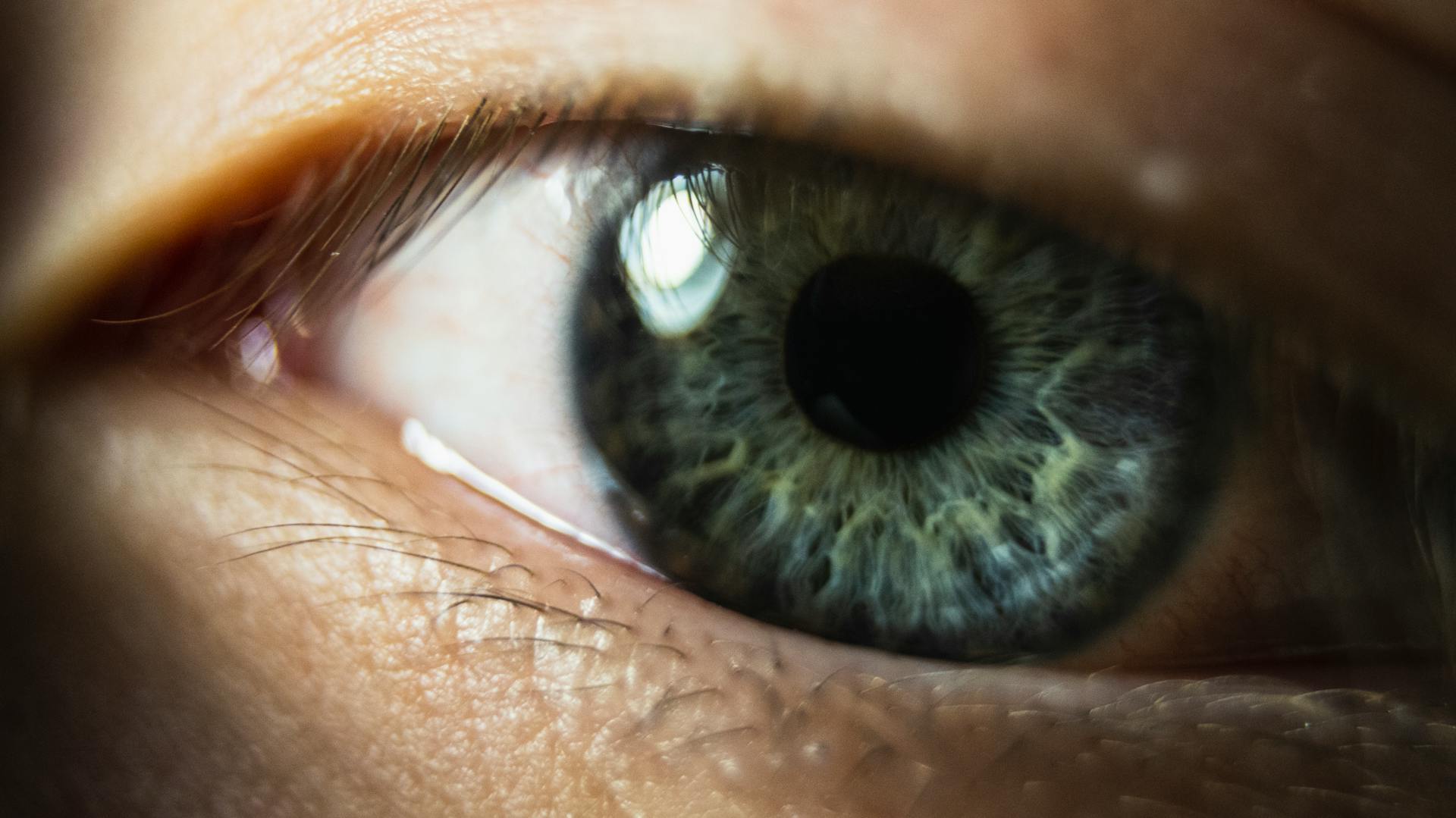
The lacrimal punctum is usually visible as a small dot on the medial canthus, the area where the upper and lower eyelids meet. In dogs with an imperforate lacrimal punctum, this dot is either missing or not visible.
This condition can be congenital, present at birth, or acquired later in life due to injury or disease.
What Is Punctum?
Imperforate lacrimal punctum in dogs is a congenital disorder.
The lacrimal punctum is located at the corner of the eye, and it plays a crucial role in draining tears.
Tears occur naturally to lubricate your dog's eyes and drain through the lacrimal punctum.
The lacrimal punctum is not just one opening, but rather two - the inferior and the superior.
Dogs have two lacrimal puncta, and imperforate lacrimal punctum can affect one or both of these openings.
Any dog can develop imperforate lacrimal punctum, but some breeds are more predisposed to it than others.
Specific breeds that are prone to imperforate lacrimal punctum include the Samoyed, American Cocker Spaniel, Poodle, Bedlington Terrier, and Golden Retriever.
Symptoms and Diagnosis in Dogs
If you notice any unusual changes to your dog's eyes, it's essential to have them checked out by your veterinarian.
Watery eyes or epiphora, rust colored staining at the corner of the eye or down the face, excessive blinking or blepharospasm, rubbing the affected eye or scratching at the eye, and swelling of the eye are all symptoms of imperforate lacrimal punctum in dogs.
If your veterinarian suspects a blockage in the tear duct, they will try to flush the nasolacrimal duct to determine if there is a blockage.
A bacterial culture test may also be done to determine if the excessive tearing is being caused by an underlying bacterial infection.
Some veterinarians may not be comfortable diagnosing eye diseases or conditions, in which case they will refer you to a specialist called a canine ophthalmologist.
A different take: Dogs Eye Swollen from Allergies
Causes and Treatment of Punctum
Imperforate lacrimal punctum in dogs is an inherited disorder, often caused by a blockage in the tear duct or a small opening that easily becomes blocked.
Some breeds, such as Cocker Spaniels, are more prone to this condition due to a hereditary defect in the formation of the nasolacrimal duct.
Tears have no place to drain when the lacrimal punctum is blocked, leading to excessive tearing and potential eye irritation.
In some cases, the obstruction is related to the shape and size of the dog's head and muzzle.
To treat imperforate lacrimal punctum, surgery is often the best option to open the lacrimal punctum and allow tears to drain properly.
Your veterinarian will discuss the best treatment option with you and may prescribe antibiotic medications to reduce the risk of infection after surgery.
Keep in mind that your dog will need regular post-surgical eye checks to ensure the lacrimal punctum remains open and functioning properly.
You might enjoy: Gastric Dilatation Volvulus Surgery
Causes of Punctum
Imperforate lacrimal punctum in dogs is an inherited disorder that occurs when the lacrimal punctum in the tear duct becomes blocked. Some dogs are born with blockages to the lacrimal punctum, while others are born with very small openings that easily become blocked.
The shape and size of a dog's head and muzzle can also cause obstruction of the lacrimal duct. This is a common issue in breeds like Cocker Spaniels.
A hereditary defect in the formation of the nasolacrimal duct can lead to imperforate puncta, where the lacrimal punctum lacks an opening to the conjunctiva. This condition often affects Cocker Spaniels, but other breeds may also be affected.
Inflammation or infection within the eye or lacrimal duct can cause swelling that blocks the duct, leading to lacrimal duct obstruction.
Expand your knowledge: Can Allergies Cause Swollen Lymph Nodes in Dogs
Treatment Options
Surgery to open the lacrimal punctum is a common treatment option for imperforate lacrimal punctum.
Many veterinarians or canine ophthalmologists will suggest this procedure to correct the blockage.
Antibiotic medications, such as Metronidazole and Enrofloxacin, are prescribed to reduce the risk of infection after surgery.
Antibacterial and anti-inflammatory medications may also be prescribed to aid in the healing process.
It's essential to follow all dosage instructions on medications prescribed by your veterinarian.
Post-surgical eye checks are usually required once a month to ensure the lacrimal punctum remains open and to check for any signs of infection.
Until the post-operative recovery period is over, it's best to keep your dog indoors to prevent debris from getting into the eye and injuring the surgical site.
An Elizabethan collar, also known as an E-collar, may be required to prevent your dog from aggravating the eye.
Recovery in Dogs
In most cases, dogs can recover from an imperforate lacrimal punctum without any long-term complications.
Dogs may exhibit discharge or crusting around the eyes, which can be a sign of tear duct obstruction.
Recovery typically takes around 2-4 weeks with proper treatment and care.
Proper treatment includes flushing the tear ducts and applying topical ointments to the affected area.
With proper care, most dogs can return to their normal activities within a few weeks.
You might enjoy: Lick Granuloma Dog Home Treatment
Frequently Asked Questions
What would happen to tear drainage if the lacrimal punctum were blocked?
If the lacrimal punctum is blocked, tears will build up and overflow onto the cheek, even when you're not crying. This can lead to excess tear flow and potentially cause eye irritation.
How do you treat punctal atresia?
Treatment for punctal atresia may involve a surgical procedure to open or enlarge the duct opening, typically performed under general anesthesia with the aid of an operating microscope. This procedure can help restore normal tear drainage and alleviate related symptoms.
Sources
- https://wagwalking.com/condition/imperforate-lacrimal-punctum
- https://www.petmoo.com/dogs/imperforate-lacrimal-punctum-in-dogs/
- https://vcahospitals.com/know-your-pet/lacrimal-duct-obstruction-in-dogs
- https://www.merckvetmanual.com/eye-diseases-and-disorders/ophthalmology/nasolacrimal-and-lacrimal-apparatus-in-animals
- https://www.msdvetmanual.com/dog-owners/eye-disorders-of-dogs/disorders-of-the-nasal-cavity-and-tear-ducts-in-dogs
Featured Images: pexels.com