A golf ball size lump on your dog's neck overnight can be alarming, but it's essential to remain calm and take action quickly. A golf ball size lump on a dog's neck is often a sign of a cyst or abscess, which is a pocket of pus that forms as a result of a bacterial infection.
The key to treating a golf ball size lump on a dog's neck overnight is to identify the underlying cause and take steps to reduce swelling and prevent infection. This can be done by applying a warm compress to the affected area to bring the pus to a head and encourage drainage.
If the lump is painful or tender to the touch, it's crucial to seek veterinary attention immediately. A golf ball size lump on a dog's neck can be a sign of a more serious underlying condition, such as a tumor or a parasite infestation.
In some cases, a golf ball size lump on a dog's neck can be caused by a skin infection, such as a staph infection, which can be treated with antibiotics.
Additional reading: American Bully Ear Infection
Causes and Symptoms
A golf ball-sized lump on your dog's neck can be alarming, but understanding the possible causes can help you identify the issue.
Salivary mucoceles can cause a round lump on the neck, and they're usually not painful unless they become large or infected. Large or infected salivary mucoceles may cause non-specific signs of illness, including lethargy and loss of appetite.
A swollen lymph node, often located in the neck area, can also form a golf ball-sized lump. Lymph nodes can swell due to upper respiratory infections or dental diseases.
A cervical abscess may also be the cause of the lump, often resulting from swallowing a foreign object that easily splinters.
For another approach, see: Chihuahua Sized Dogs
Symptoms of Salivary Mucoceles
A salivary mucocele can affect various glands or associated ducts and usually looks and feels like a round lump.
These swollen areas are not typically painful in the early stages unless they become large enough to put pressure on another part of the anatomy.
Large or infected salivary mucoceles may cause dogs to show non-specific signs of illness, including lethargy and loss of appetite.
The specific signs of a salivary mucocele will depend on the type, with four types of sialoceles, each named for the location where they occur.
Causes of Salivary Mucoceles in Dogs
Salivary mucoceles in dogs can be caused by a variety of factors. Traumatic injuries to the salivary glands and ducts are a common cause, often resulting from oral injuries like chewing on sticks or bite wounds from other animals.
Oral injuries can range from minor to severe, and even pulling on a choke chain or prong collar can cause a neck injury that leads to a salivary mucocele.
Sialolithiasis, a rare condition in dogs, can also cause stones to form in the salivary glands or ducts, leading to a blockage and rupture that results in a salivary mucocele.
Foreign bodies, such as grass awns, can block the salivary glands and lead to rupture, causing a salivary mucocele.
Any dog breed can develop sialoceles, but some breeds are more often affected, including German shepherds, dachshunds, poodles, greyhounds, and Australian silky terriers.
Here are some common types of injuries that can lead to salivary mucoceles:
- Oral injury from chewing on an object, such as a stick
- Bite wounds from another animal
- Neck injury from pulling on a choke chain or prong collar
Diagnosis and Treatment
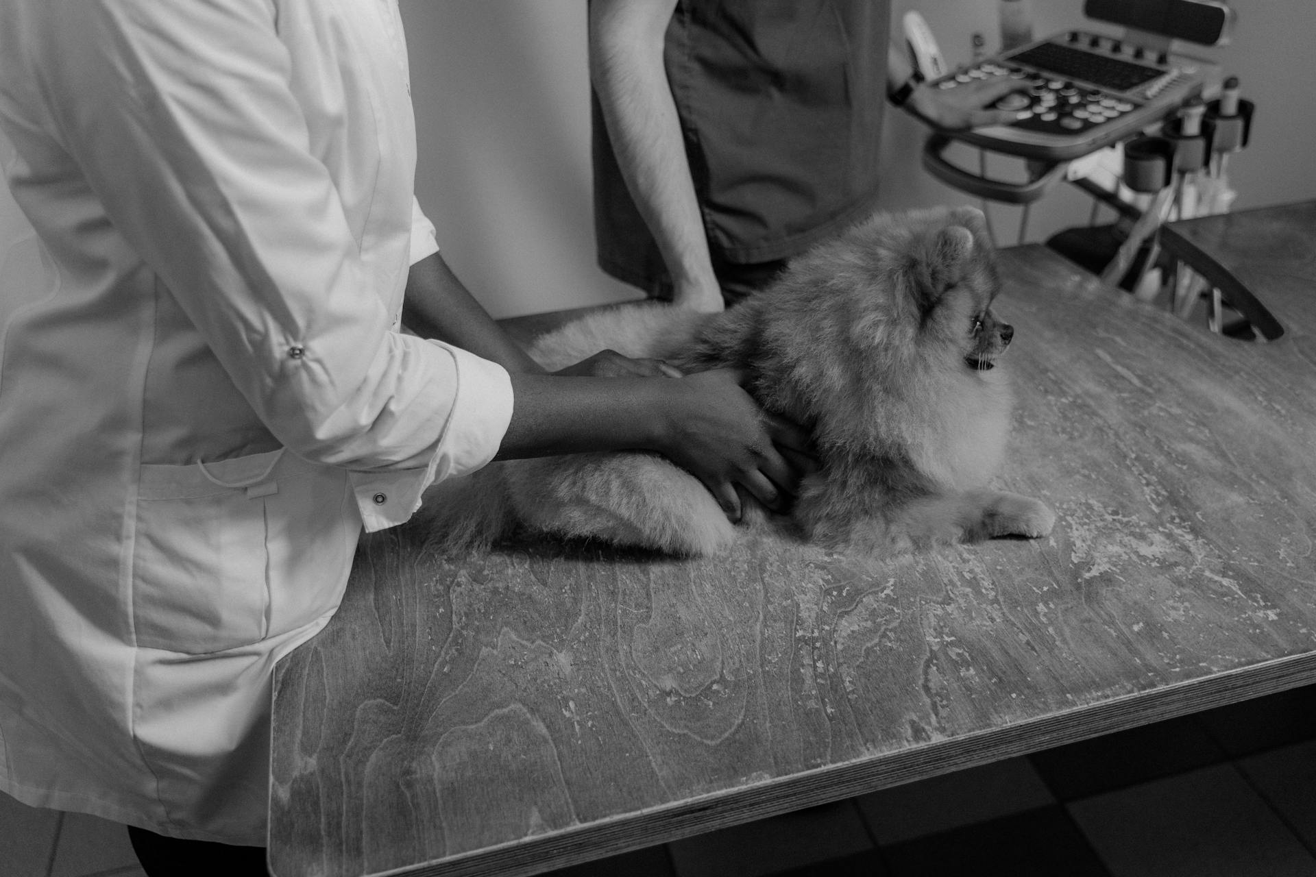
Diagnosis involves a physical examination and a sample collection. Your veterinarian will look closely at the swollen area and may collect a sample to make a definitive diagnosis.
A fine needle aspirate is often used to take a sample from the lump. This involves puncturing the lump with a needle and sucking out some cells to examine under a microscope.
The sample may be sent to a lab for analysis to confirm the diagnosis. A veterinary pathologist will analyze the fluid to determine what kinds of cells are present and confirm whether or not the swelling is a sialocele.
Treatment typically requires draining and surgical intervention. Draining may be used to offer temporary relief until surgery can be performed, but it's not a recommended long-term solution as it can lead to inflammation or infection.
A definitive treatment involves surgical removal of the affected salivary glands and associated ducts. This is a delicate procedure that is typically performed by a board-certified veterinary surgeon.
You might enjoy: My Dogs Not Eating and Is Lethargic
How Vets Diagnose Salivary Mucoceles in Dogs
Diagnosing a salivary mucocele in dogs involves a thorough examination by your veterinarian. They will start by discussing your pet's history to understand the symptoms and when they first appeared.
Your veterinarian will then perform a physical examination, closely looking at the swollen area. They may want to collect a sample to make a definitive diagnosis.
The diagnosis process involves three steps: aspiration, visual inspection, and lab analysis. Aspiration involves using a needle and syringe to collect fluid for testing, which may require sedation depending on the location.
The fluid from a sialocele is generally clear, yellowish, or blood-tinged in color and slightly viscous like saliva. Your veterinarian may be able to see right away that it is saliva, but will likely send the fluid to a lab for analysis to be certain.
Lab analysis involves a veterinary pathologist examining the fluid to determine what kinds of cells are present and confirm whether or not the swelling is a sialocele. This analysis can also rule out infections, cancer, and other potential causes for the swelling.
A fresh viewpoint: Female Dog Leaking Smelly Fluid
Here are the three steps involved in diagnosing a salivary mucocele:
- Aspiration: Your vet may use a needle and syringe to collect fluid for testing.
- Visual Inspection: The fluid from a sialocele is generally clear, yellowish, or blood-tinged in color and slightly viscous like saliva.
- Lab Analysis: A veterinary pathologist will analyze the fluid to determine what kinds of cells are present.
Treating Salivary Mucoceles in Dogs
If your dog has a salivary mucocele, it's essential to seek veterinary attention as soon as possible. Without treatment, a salivary mucocele can become infected and abscessed.
Your vet may recommend draining the mucocele to provide temporary relief until surgery can be performed. However, most sialoceles will eventually recur after being drained, and continued draining is not recommended as it can lead to inflammation or infection.
Draining is not a long-term solution, but rather a temporary fix to alleviate symptoms. The definitive treatment of a salivary mucocele involves surgical removal of the affected salivary glands and associated ducts.
Surgery is typically performed by a board-certified veterinary surgeon and may involve placing drains at the surgical site to prevent new fluid accumulation. This is a delicate procedure that requires great care and attention.
Here are the treatment options for salivary mucoceles:
- Draining: temporary relief until surgery can be performed
- Surgery: removal of affected salivary glands and associated ducts
It's crucial to work closely with your vet to determine the best course of treatment for your dog's specific case.
Prognosis and Expectations
If your dog has a salivary mucocele, the prognosis is generally good, with most dogs recovering well from surgery.
The surgical removal of the affected salivary gland is usually a successful procedure, and complications are rare.
Your veterinarian will provide you with post-operative care instructions, which you should follow carefully.
These instructions may include giving your dog medications as directed and keeping the incision, drain sites, and any bandages clean and dry.
Follow-up visits with your veterinarian will be necessary to ensure your dog's recovery is progressing smoothly.
Here's a brief overview of what to expect during the recovery period:
Lump Types and Swelling
A golf ball-sized lump on your dog's neck can be a cause for concern, and it's essential to identify the possible causes. Lymph nodes are located in various parts of the dog's body, and the neck is one area where the lymph node could be felt, referred to as submandibular lymph nodes.
The lump may have formed because these lymph nodes are affected by swelling, which can be caused by upper respiratory infection or dental diseases. It's also possible that the lump could be a sign of something cancerous like lymphoma.
Explore further: Why Are My Dog's Balls Peeling?
In some cases, the lump may be a cervical abscess, which typically happens after a dog swallows a stick or other foreign object that easily splinters. Abscesses are usually firm to touch and may cause redness around the dog's neck area.
Here's a summary of possible lump types and swelling:
It's essential to have your veterinarian check the lump as soon as possible to determine the cause and provide the best course of treatment.
Dog Lymph Node Swelling
Dog lymph node swelling can be a cause for concern. Lymph nodes are located in various parts of a dog's body, including the neck, where the submandibular lymph nodes can be felt.
A swollen lymph node can form a golf-ball sized lump. This lump may be due to various reasons, including upper respiratory infection and dental diseases.
If your dog has a soft, moveable, and golf-ball sized lump on the back of their leg, it could be a fatty tumor or lipoma. It's also possible that it's a swollen lymph node near the knee.
On a similar theme: Golf Balls Safe
It's essential to have your veterinarian check the lump as soon as possible. A needle biopsy may be performed to obtain a sample of cells from the lump. This can help determine if the mass is cancerous.
Removing a mass on a leg while it's small is generally safer and less expensive. It's also easier to close the incision with enough skin remaining.
Dogs with Cervical Abscess
A golf ball-sized lump on your dog's neck could be a cervical abscess, typically caused by swallowing a stick or other foreign object that easily splinters.
This type of abscess is usually firm to the touch and may cause redness around the dog's neck area.
It's crucial not to try to drain the abscess yourself, as this can open up the opportunity for bacteria to enter your dog's body.
The best course of action is to have your dog checked by a vet as soon as possible, as they can provide further diagnostics and treatment.
Benign Lumps
Benign lumps are a common occurrence in dogs, and it's essential to know what to look out for. A histiocytoma is a type of benign growth that can go away on its own, especially in younger dogs. It's often raised, hairless, and sometimes ulcerated.
If you suspect your pet has a lump, don't hesitate to contact your vet directly for an assessment. They can provide you with more information and a care plan.
Some common types of benign lumps include histiocytomas, lipomas, apocrine gland cysts, and melanocytomas. Lipomas are abnormal growths of fatty tissue that don't spread but can get quite large.
Here are some key characteristics of benign lumps:
- Histiocytoma: raised, hairless, sometimes ulcerated
- Lipoma: abnormal growth of fatty tissue, can get quite large
- Apocrine Gland Cysts: originate from glands in the skin, often on the head and neck
- Melanocytoma: darkly pigmented, typically small, seen on the head and forelimbs of middle-aged or older dogs
Melanocytomas arise from melanin-producing cells and are usually small. If you notice any changes in your dog's lump, such as growth or discomfort, consult your vet immediately.
Golf Ball Sized Lump on Dog
A golf ball sized lump on your dog can be a cause for concern.
If the lump is on the neck, it could be a swollen lymph node, which can be a sign of an upper respiratory infection or dental disease.
Swollen lymph nodes can be felt in various parts of the body, including the neck, where they are referred to as submandibular lymph nodes.
It's essential to have your dog checked by a veterinarian as soon as possible, especially if the lump is golf ball-sized, as it could be a sign of something cancerous like lymphoma.
A soft golf ball size mass on a leg may be a fatty tumor or lipoma, but it could also be a swollen lymph node near the knee.
Removing a mass on a leg while it's small is generally safer and less expensive than waiting until it grows.
If your dog has a swollen area on their neck, your veterinarian may perform a physical examination and collect a sample for testing.
There are three steps to diagnose a salivary mucocele in dogs: aspiration, visual inspection, and lab analysis.
Here are the three steps to diagnose a salivary mucocele in dogs:
A golf ball-sized lump on the back of a dog's leg could be a cervical abscess, which typically happens after a dog swallows a foreign object that easily splinters.
What to Do
If your dog has a lump on his neck, it's essential to take immediate action.
Call the vet as soon as possible. They may ask you to bring the dog in for further diagnostics.
A golf ball-sized lump could be a sign of something serious, like lymphoma, so it's best to have the dog checked as soon as possible.
Frequently Asked Questions
Can a cancer lump appear overnight on a dog?
Cancer lumps can appear rapidly in dogs, but it's also possible for them to develop slowly over time, making early detection challenging. If you suspect a lump on your dog, consult a veterinarian for a proper evaluation and diagnosis
Can a lipoma show up overnight dog?
Lipomas are typically slow-growing tumors, so it's unlikely for a new mass to appear overnight. If you notice a sudden growth, it's worth investigating further to determine its cause.
Sources
- https://www.thesprucepets.com/sialocele-salivary-mucocele-3384320
- https://www.mypetchild.com/dogs/health/golf-ball-sized-lump-neck/
- https://www.dogster.com/lifestyle/lumps-on-dogs
- https://www.vetinfo.com/vets/answers/golf-ball-sized-lump-on-dog-that-moveable-and-soft
- https://www.vmamodesto.com/blog/lumps-bumps-mass-dog/
Featured Images: pexels.com