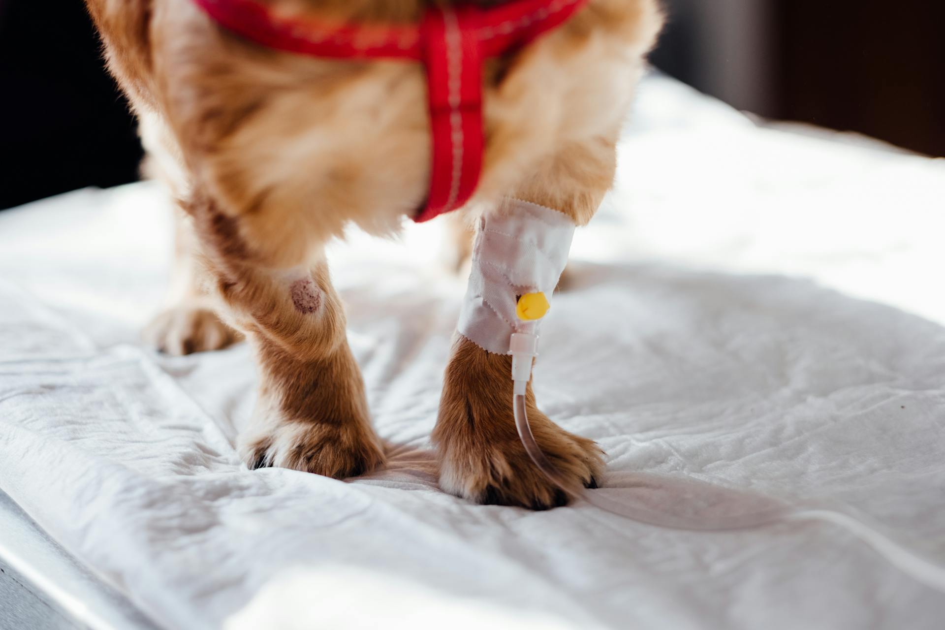
Subcutaneous fluid dog lumps can be a concerning sight for any dog owner. They are typically found just beneath the skin and can feel like a soft, squishy mass.
These lumps are usually filled with a type of fluid called serous fluid, which is a clear, watery liquid. This fluid can accumulate due to various reasons such as trauma, infection, or even a blockage in the lymphatic system.
The size and location of the lump can vary greatly, and it's not uncommon for them to appear suddenly. In some cases, the lump may be tender to the touch, while in others it may be completely painless.
It's essential to have your dog checked by a veterinarian as soon as possible to determine the cause and best course of action.
Take a look at this: Fluid Lump on Dog Chest
Causes and Types
Subcutaneous fluid dog lumps can be caused by a variety of factors, including infections, allergies, and trauma.
Infections can lead to the formation of abscesses or cellulitis, which can cause lumps under the skin.
Allergies can cause hives, itching, and swelling, which may present as a lump.
Trauma, such as a bite or scratch, can cause swelling and bruising, leading to a lump.
Some common types of subcutaneous fluid dog lumps include abscesses, cysts, and hematomas.
Abscesses are painful, fluid-filled pockets that can become infected and cause a lump.
Cysts are non-cancerous, fluid-filled sacs that can be caused by a variety of factors, including genetics and trauma.
Hematomas are collections of blood that can form after trauma, leading to a lump.
It's essential to consult a veterinarian to determine the cause and type of subcutaneous fluid dog lump.
A different take: Large Fluid-filled Lump on Dog Stomach
Diagnosis and Testing
Veterinarians use fine needle aspiration (FNA) to determine the cause of a lump by removing cells or fluid from the lump, which is then analyzed under a microscope.
This test can help rule out causes or guide further diagnostic testing, but it may not always give an exact answer.
During a biopsy, the entire lump or a small section of the lump is surgically removed, and the tissue is submitted to a veterinary pathologist for examination.
A biopsy is often necessary for a more specific diagnosis, especially if the lump is large or has been present for a long time.
To ensure a representative cytologic sample, veterinarians must acquire a satisfactory sample, prepare it correctly, and screen for cellularity.
Inadequately cellular samples can be a problem, occurring in up to 16.8% of cases.
Detailed descriptions of how to obtain cytologic samples via FNA are available elsewhere.
To provide accurate cytologic interpretation, veterinarians need to know accurate and detailed patient history and mass characteristics.
A full patient signalment, including age, sex, and breed, is essential for cytologic interpretation.
Patient lifestyle and travel history, as well as specifics about the lesion, should also be included.
Any relevant diagnostic test results or clinical information should be provided to the pathologist.
Here are the essential details to include when submitting slides to a pathologist:
- Full patient signalment (e.g., age, sex, castration or reproductive status, species, breed)
- Patient lifestyle/travel history
- Specifics about the lesion (e.g., duration, location, distribution, size, gross description)
- Any other relevant diagnostic test results or clinical information
When to Worry
If the lump persists for more than a few weeks with no change in size, you should consult your veterinarian.
A painful lump is a clear sign that something is amiss, and you should seek veterinary attention right away.
Lumps that start oozing or draining pus are a serious concern and require immediate veterinary care.
A growing lump is a cause for concern, and it's essential to have it checked by a veterinarian to rule out any underlying issues.
Any lump that doesn't seem to be going away on its own should be evaluated by a veterinarian to ensure it's not a sign of something more serious.
Frequently Asked Questions
What happens if a dog gets too much subcutaneous fluid?
Administering too much subcutaneous fluid to a dog can lead to pulmonary or interstitial edema, a potentially life-threatening condition. It's essential to follow the recommended fluid administration guidelines to avoid complications
How long does it take subcutaneous fluids to absorb a dog?
The absorption time for subcutaneous fluids in dogs can vary from a few minutes to several hours, depending on their hydration status. If not absorbed, fluids may be drawn to the lower abdomen or legs, so wait for the next administration time before giving additional fluids.
Can subcutaneous fluids leak out of a dog?
Yes, subcutaneous fluids can leak out of a dog, appearing as a clear or blood-tinged fluid at the injection site. If this occurs, gentle pressure can help stop the flow.
Sources
- https://www.dogster.com/ask-the-vet/lump-after-subcutaneous-injection-dog
- https://fairhavenvet.com/subcutaneous-fluids-how-to/
- https://todaysveterinarypractice.com/clinical-pathology/small-animal-skin-cytology/
- https://www.dogcancer.com/perspectives/diagnosis-and-medical-procedures/staying-vigilant-with-mass-aspirates/
- https://www.smalldoorvet.com/learning-center/medical/mast-cell-tumors-mastocytomas/
Featured Images: pexels.com


