Histiocytoma in dogs is a common skin condition that affects many breeds, but its exact cause remains a mystery.
The condition is most often seen in young dogs, typically between the ages of 6 months to 2 years old.
Histiocytoma is a benign tumor made up of histiocytes, a type of immune cell that plays a key role in the body's defense against infection.
It usually appears as a small, firm, and painless lump on the skin, often on the legs or face.
For more insights, see: What Is Histiocytoma in Dogs
Causes and Pathogenesis
The exact cause of histiocytoma in dogs is still unknown, but research suggests that it may be linked to dysregulation of the immune system, specifically the proliferation, activation, and function of dendritic cells and T cells after antigenic stimulation.
This theory is supported by the fact that some cases of histiocytosis, including cutaneous and systemic histiocytosis, respond to immunosuppressive therapy.
Dysregulation of the immune system can lead to a range of symptoms, including the formation of granulomatous inflammation, which is characterized by large, pale, round to oval histiocytes with indented vesicular nuclei.
These histiocytes are thought to be of activated dermal (interstitial) dendritic cell origin, given their expression of Thy1 and CD4 surface markers.
In some cases, the immune system's response to an unknown antigen may lead to a secondary phenomenon, where T cells infiltrate the affected area, potentially attracting and retaining migrating dendritic cells.
Canine Cutaneous Tumors
Canine cutaneous tumors, also known as histiocytomas, can occur in dogs of any age, but the incidence drops significantly after 3 years of age.
Some breeds are more prone to histiocytomas, including boxers, dachshunds, cocker spaniels, Great Danes, Shetland sheepdogs, and bull terriers.
Histiocytomas are usually small, fast-growing, raised, and hairless lesions that are typically less than 2-3 cm in size.
These tumors most commonly arise on the extremities, head, ears, or neck, and are usually solitary, but some dogs may have multiple lesions at diagnosis.
Despite their rapid growth, histiocytomas are considered benign, meaning they are not cancerous.
Pathogenesis

The cause of both cutaneous and systemic histiocytosis in dogs is still unknown, but a prevailing hypothesis suggests that it's linked to dysregulation of dendritic cells and T cells after antigenic stimulation.
This theory is supported by the fact that some cases respond to immunosuppressive therapy, which suggests that the immune system is playing a key role in the development of the disease.
The specific antigenic process leading to the immune dysregulation hasn't been identified yet, but it's clear that it's a complex interplay of multiple factors.
In both cutaneous and systemic histiocytosis, the histologic features are identical, with biopsy specimens showing numerous large, pale, round to oval histiocytes with indented vesicular nuclei.
These histiocytes express specific surface markers, such as CD1b, CD1c, CD11c, and MHC II, which are indicative of their derivation from dendritic antigen-presenting cells.
The lesions in both types of histiocytosis tend to wax and wane, with spontaneous regression in the early stages of disease, but this is often followed by the development of new lesions in other locations.
This pattern of disease progression suggests that the immune system is still actively involved, even after the initial lesions have regressed.
Symptoms and Treatment
Histiocytomas in dogs are typically small, solitary, hairless lumps that appear as a small, solitary, hairless lump, most commonly found on the head, neck, ears, and limbs.
They are usually less than 2.5 cm in diameter and often ulcerate. Shar Peis may be predisposed to multiple histiocytomas.
Diagnosis is made through cytology of the mass, which reveals cells with clear to lightly basophilic cytoplasm and round or indented nuclei with fine chromatin and indistinct nucleoli.
Intriguing read: Merrick Dog Food for Small Dogs
Symptoms
Histiocytomas in dogs are often found in young dogs and appear as small, solitary, hairless lumps.
These lumps are usually less than 2.5 cm in diameter and can be found on the head, neck, ears, and limbs.
Ulceration of the mass is common, which can be a concern for dog owners.
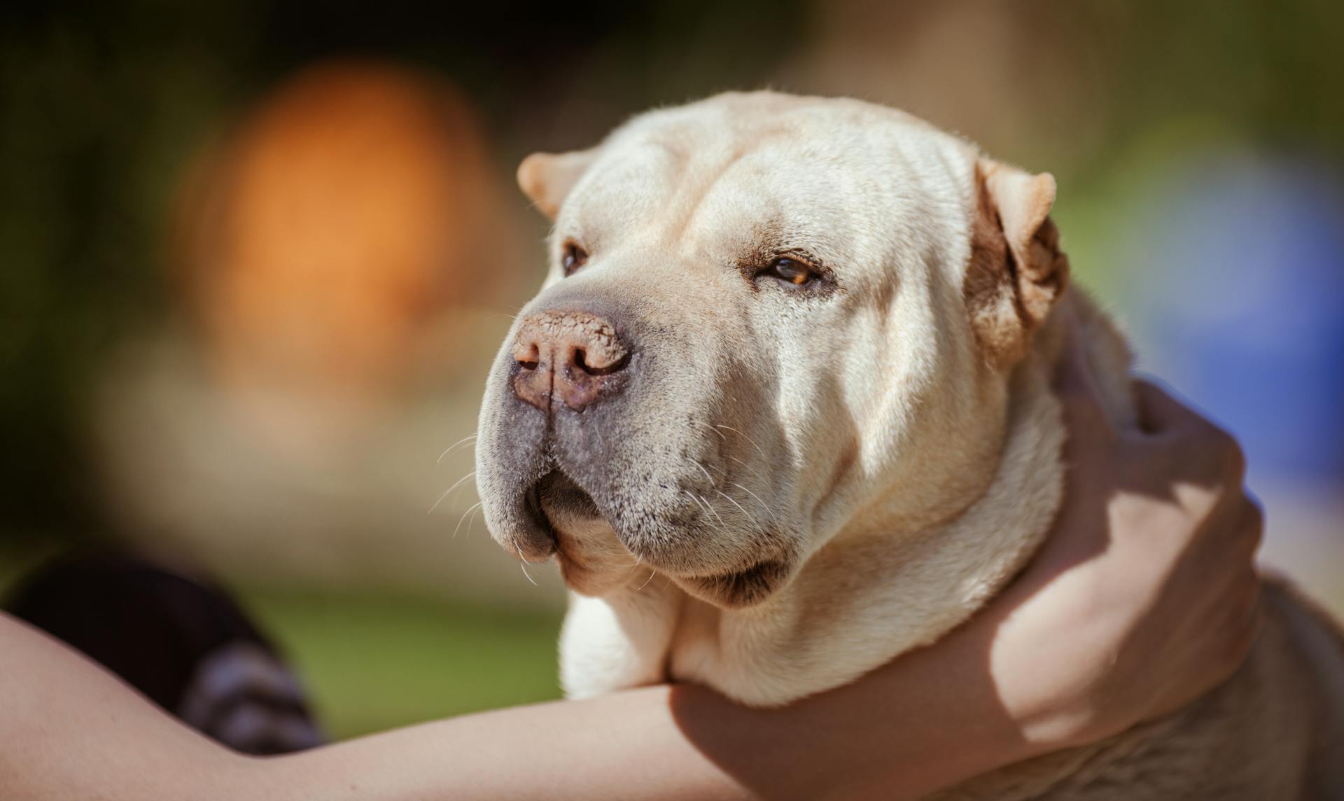
Shar Peis may be predisposed to multiple histiocytomas, making them more susceptible to this condition.
Cytology of the mass reveals cells with clear to lightly basophilic cytoplasm and round or indented nuclei with fine chromatin and indistinct nucleoli.
Treatment and Prognosis
In most cases, histiocytomas spontaneously regress within three months.
Some people might be worried about the possibility of metastases to the lymph nodes, but it's worth noting that this has rarely been reported.
The behavior of histiocytomas can be unpredictable, and in some cases, it can resemble a more aggressive disease called Langerhans cell histiocytosis.

Dogs with multiple histiocytomas can experience a protracted clinical course, with new nodules forming while others regress.
Surgical excision is often curative, and it's recommended for patients with nodules that are ulcerated, infected, or pruritic.
In general, adjunct therapy like corticosteroids is unnecessary, and the prognosis for solitary regressing cutaneous histiocytoma is excellent.
Even in cases where multiple nodules are present, regression is ultimately expected, and a good prognosis can be given.
Frequently Asked Questions
How do you prevent histiocytomas in dogs?
Unfortunately, there is no known way to prevent histiocytomas in dogs. Regular veterinary check-ups can help identify any potential skin issues early on.
Is histiocytoma in dogs contagious?
No, histiocytomas in dogs are not contagious to humans or other animals. They are a common skin condition in young dogs, typically under 2 years old.
What is the healing process of histiocytoma?
Histiocytomas typically heal within 3 months, with surgical cases requiring a 2-week recovery period and post-operative care to prevent complications.
Sources
- https://www.mspca.org/angell_services/color-atlas-canine-cutaneous-round-cell-tumors/
- https://www.dvm360.com/view/overview-canine-histiocytic-disorders
- https://vcahospitals.com/know-your-pet/skin-cutaneous-histiocytoma
- https://en.wikipedia.org/wiki/Histiocytoma_(dog)
- https://www.marvistavet.com/histiocytoma.pml
Featured Images: pexels.com


