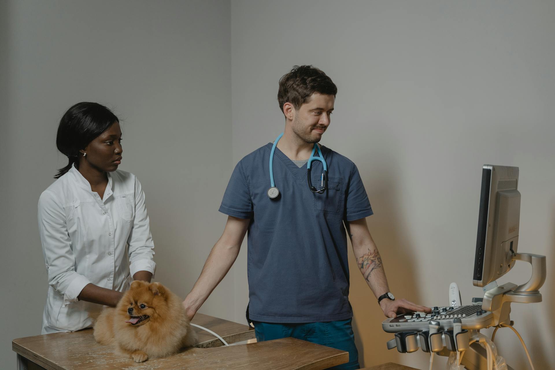
Understanding canine abdomen anatomy is crucial for identifying potential health issues through ultrasound imaging. The abdominal cavity of a dog contains several organs, including the stomach, small intestine, and liver.
The stomach is a muscular sac that can hold up to 2 liters of food and digestive fluids. It's divided into four sections: the fundus, body, antrum, and pylorus.
The small intestine is a long, thin tube where most of the nutrient absorption takes place. It's divided into three parts: the duodenum, jejunum, and ileum.
The liver is a vital organ responsible for detoxification, metabolism, and production of bile. It's located on the right side of the abdominal cavity and is divided into lobes.
Canine Abdomen Anatomy
The canine abdomen is a complex and fascinating area of study. The xiphoid process is a key landmark in this region, and it's located at the bottom of the sternum, just above the abdomen.
To begin a tour of the abdomen, the dog should be positioned in dorsal or lateral recumbency, allowing for easy access to the area. This makes it easier to perform procedures and exams.
The probe should be placed just caudal to the xiphoid process, marking the starting point for the examination. This position provides a clear view of the abdominal contents.
The abdominal cavity is a vital space that houses many essential organs, including the stomach, intestines, liver, and kidneys.
Ultrasound Imaging
Ultrasound imaging is a valuable tool for evaluating canine abdomen anatomy. The liver is a key organ to examine, and it should be visualized in both long and short axes to get a complete picture.
The liver has a coarse echogenicity and contains vessels, including the portal veins and hepatic veins. The portal veins have an outer hyperechoic wall, while the hepatic veins are hypoechoic tubular structures without hyperechoic walls.
To evaluate the liver, use a non-distance motion and angle the probe cranially to view the midsection of the liver. Depending on liver size, use a non-distance motion and angle the probe to the left or right to view the left or right side of the liver.
Here are some key points to keep in mind when evaluating the liver:
- The normal liver has a coarse echogenicity and contains vessels.
- The portal veins have an outer hyperechoic wall.
- The hepatic veins are hypoechoic tubular structures without hyperechoic walls.
In addition to the liver, it's also important to evaluate the stomach, left kidney, and urinary bladder in both long and short axes. The stomach can be visualized in short axis by sliding the transducer caudally from the xiphoid position. The left kidney and left adrenal gland can be identified by moving the transducer with a medial and slightly caudal distance motion from the level of the spleen.
Explore further: Canine Kidney Cancer Symptoms
Echogenicity & Echotexture
Echogenicity is a challenging aspect to evaluate in ultrasound imaging, as it's easy to adjust the gain too high or too low, resulting in images that are hyperechoic or hypoechoic overall.
The echotexture of an organ or structure is also crucial, and it's evaluated in addition to overall gray scale or echogenicity.
The echotexture can vary greatly between different organs and structures, and it's essential to understand what's normal for each one.
Evaluating echogenicity and echotexture requires a good understanding of imaging optimization, as excessive gain can lead to poor-quality images.
It's essential to strike the right balance when adjusting the gain to obtain high-quality scans.
Imaging Planes
Imaging Planes are crucial in ultrasound evaluation, and it's essential to use 2 different planes for each organ, especially when abnormalities are suspected.
You might be wondering why 2 planes are necessary. The imaging planes used in organ evaluation often don't align with standard imaging planes, so an oblique imaging plane is often used to best visualize an organ.
To ensure accuracy, make sure all images are labeled appropriately, with correct orientation. These images will become an official part of the patient's medical record, so it's essential to get it right.
When evaluating an organ, it's not uncommon to use an oblique imaging plane. This allows you to get a clear view of the organ, even if it's not in a standard position.
To label images correctly, you need to consider the orientation of the image. This can be tricky, but it's essential to get it right to avoid confusion.
Specific Organ Ultrasound
Ultrasound is a valuable tool for visualizing the abdominal organs of our canine friends. The liver, in particular, is a key organ to examine, and it's relatively easy to do so with a little practice.
To view the liver, angle the probe cranially and use a non-distance motion to bring the midsection into view. Depending on the liver size, you may need to adjust the angle to see the left or right side, including the gallbladder and porta hepatis.
Additional reading: Canine Liver Cancer Symptoms
When examining the liver, look for the coarse echogenicity and the presence of vessels. The portal veins are typically the dominant vessels, with an outer hyperechoic wall, while the hepatic veins are hypoechoic tubular structures without hyperechoic walls.
Here are some key differences to keep in mind when examining the liver in dogs and cats:
- Dogs: The hepatic veins enter the caudal vena cava in the dorsal right liver.
- Cats: The cystic duct and bile duct may be visualized and normally measure 2 to 3 mm in diameter.
- Cats: Fat within the falciform ligament is seen in the near field and can be isoechoic to the liver.
With practice and experience, you'll become more comfortable navigating the abdominal organs of your canine patients and using ultrasound to guide your examination.
Large Intestine
The large intestine is a crucial part of the digestive system, playing a key role in absorbing water and electrolytes from the waste material that enters it from the small intestine.
It courses between the small intestine and the anus, making it the terminal portion of the intestinal tract.
The large intestine is made up of four main parts: the cecum, colon, rectum, and anal canal.
Its average length in the dog is 0.6 meters, or 2 feet, which is shorter than the small intestine.
The large intestine is responsible for absorbing water and electrolytes, helping to form solid waste that can be eliminated from the body.
Take a look at this: Why Are Chihuahuas so Small
Liver Ultrasound
The liver is a vital organ that plays a crucial role in detoxification and metabolism. To evaluate the liver using ultrasound, it's essential to position the probe correctly.
The liver should be viewed in a non-distance motion with the probe angled cranially to bring the midsection into view. Depending on liver size, the probe can be angled to the left to view the left side of the liver or the right to view the gallbladder and right side of the liver.
The normal liver has a coarse echogenicity and contains vessels, including the portal veins with a hyperechoic wall and the hepatic veins as hypoechoic tubular structures.
The hepatic veins can be seen as they taper toward the periphery of the liver and enlarge centrally within the liver. They enter into the caudal vena cava in the dorsal right liver.
The hepatic artery branches and intrahepatic bile ducts are not visualized normally. However, in cats, the cystic duct and bile duct may be visualized and normally measure 2 to 3 mm in diameter.
Fat within the falciform ligament can be seen in the near field, particularly in cats, and can be isoechoic to the liver.
Here are some key points to keep in mind when evaluating the liver using ultrasound:
- Use a non-distance motion to view the liver
- Angle the probe to view the left or right side of the liver
- Look for the portal veins with a hyperechoic wall and the hepatic veins as hypoechoic tubular structures
- Note the size and location of the hepatic veins
- Be aware that the hepatic artery branches and intrahepatic bile ducts are not visualized normally
- Check for fat within the falciform ligament, especially in cats.
Stomach Ultrasound
When performing an ultrasound of the stomach, it's essential to start from the xiphoid position and slide the transducer caudally to visualize the stomach in short axis. This allows you to evaluate the stomach in its entirety in both long- and short-axis images.
The pyloroduodenal junction is a critical area to visualize, and it appears as a thickened area of the muscularis between the pylorus and proximal duodenum when the transducer is swept toward the right side.
In felines, the submucosa of the stomach is often hyperechoic due to fat deposition in this layer, which can be a notable feature to observe during the ultrasound.
Readers also liked: Anatomy of Canine Stomach
Ultrasound of Left Kidney & Adrenal Gland
Ultrasound of the Left Kidney & Adrenal Gland is a crucial imaging technique for evaluating the left kidney and adrenal gland. The procedure starts by moving the transducer with a medial and slightly caudal distance motion from the level of the spleen to identify the left kidney.
Broaden your view: Canine Salivary Gland Anatomy
To visualize the left kidney, use long and short axes imaging planes, as shown in Figure 10. The left kidney can be seen in both sagittal and transverse views.
The abdominal aorta is seen in long axis when the probe is angled medially from the left kidney, as demonstrated in Figure 11. This allows for a clear visualization of the abdominal aorta.
The celiac and cranial mesenteric arteries, along with the left renal artery, form the vascular delimiters for the area where the left adrenal gland can be found, as shown in Figure 12.
Discover more: Canine Kidney Anatomy
Ultrasound of Left Ovary
The left ovary is located caudal to the left kidney in the near field. This is a key finding in ultrasound imaging.
In an intact female, the left ovary can be indistinguishable from the surrounding fat during certain stages of estrus. This is known as anestrus.
During estrus, the ovary appears as a hypoechoic structure with multiple anechoic cysts. This is a characteristic ultrasound finding that can help identify the ovary.
The transducer should be placed just caudal to the left kidney to obtain a clear image of the ovary. This is a crucial step in the ultrasound procedure.
Urinary Bladder Ultrasound
Ultrasound of the urinary bladder is a crucial diagnostic tool for veterinarians.
To perform an ultrasound of the urinary bladder, move the transducer caudally to a central and caudal abdominal position.
Evaluate the urinary bladder in long and short axes, as shown in Figure 14, which displays a long-axis view of the urinary bladder in a dog.
The trigone area is particularly important to evaluate, especially as it extends caudally into the urethra or prostate gland.
In males, the prostate gland is visible in the long-axis image, and it's essential to note its size and shape, as seen in Figure 15.
In neutered males, the prostate gland appears as a hypoechoic fusiform-shaped enlargement of the proximal urethra.
In intact males with benign prostatic hyperplasia, the prostate gland is enlarged and hyperechoic.
Check this out: Tail Gland Hyperplasia Female Dog
Additional Views and Quality Control
Additional views can help ensure accurate diagnosis and provide a more complete understanding of canine abdomen anatomy.
To determine if a technique is appropriate for quality control, consider the exposure of abdominal viscera and the contrast between air and the patient.
Proper positioning is crucial for accurate diagnosis, and there are specific guidelines for each type of projection.
For lateral, ventrodorsal, and dorsoventral projections, the images should extend from the cranial margin of the liver to the caudal aspect of the abdomen.
Here's a quick reference guide for proper positioning:
- Lateral projections: use superimposition of the transverse processes throughout the lumbar spine to determine if a patient is in a true lateral position.
- Ventrodorsal projections: each caudal thoracic and lumbar spinous process is viewed end-on and has a distinct diamond or tear-dropped shape.
- Dorsoventral projections: positioning is similar to the ventrodorsal projection, but the pelvis will be oblique.
Additional Abdominal Views
Imaging planes are crucial for organ evaluation, and for each organ, two different imaging planes must be used, especially when suspecting abnormalities.
These imaging planes may not align with standard planes like sagittal, transverse, or dorsal planes, and an oblique imaging plane is often used to best visualize an organ.
Make sure all images are labeled correctly with the correct orientation, as these images will become an official part of the patient's medical record.
To get a better view of the urethra, especially for male dogs, a lateral view with pelvic limbs pulled cranially is obtained in addition to routine radiographs.
Pull the pelvic limbs cranially so the stifle joints are touching the ventral abdomen, and use a sandbag to keep the legs in position.
The collimation should include the cranial part from the iliac crest to the caudal-most skin margin at the ischiatic tuberosity or perineum.
Image Quality Control
Image Quality Control is a crucial step in ensuring that diagnostic images are accurate and reliable. To determine if the technique is appropriate, check if all portions of the abdominal viscera are adequately exposed with sharp contrast between the air and the patient.
The anatomy in the image should be correct, with anatomic overlap if the dog is split into cranial and caudal images. This means that the image should include the caudal sternum and xiphoid process on the lateral images or the lateral aspect of the ribs and body wall on the ventrodorsal view.

For lateral projections, use superimposition of the transverse processes throughout the lumbar spine to determine if a patient is in a true lateral position. The transverse processes appear as Nike swooshes and should be superimposed over each other.
To check patient positioning, use the following criteria:
- Lateral projections: superimposition of the transverse processes and the wings of the ilia.
- Ventrodorsal projections: each caudal thoracic and lumbar spinous process viewed end-on and having a distinct diamond or tear-dropped shape.
- Dorsoventral projections: positioning similar to the ventrodorsal projection, but with the pelvis being oblique.
Sources
- https://pubmed.ncbi.nlm.nih.gov/16766214/
- https://easy-anatomy.com/canine-digestive-system/
- https://todaysveterinarypractice.com/radiology-imaging/imaging-essentialssmall-animal-abdominal-ultrasonographya-tour-abdomen-part-1/
- https://todaysveterinarypractice.com/radiology-imaging/small-animal-abdominal-radiography/
- https://www.imaios.com/en/vet-anatomy/dog/dog-abdomen-pelvis
Featured Images: pexels.com


