The canine carpal anatomy is a fascinating topic that's essential to understand for any dog owner or enthusiast. The carpal bones are the wrist bones of a dog's front legs.
These bones are divided into three rows: proximal, intermediate, and distal. The proximal row consists of four bones: the radial, intermediate, ulnar, and accessory carpal bones.
The radial carpal bone is the most medial bone in the proximal row, and it's located on the thumb side of the paw. It plays a crucial role in supporting the weight of the dog's body.
The interosseous ligaments connect the carpal bones, providing stability and support to the wrist joint.
Canine Carpal Anatomy
The carpal bones in a dog's front paw are made up of seven bones that work together to support the dog's weight and allow for movement. These bones are the scaphoid, lunate, triquetrum, pisiform, trapezium, trapezoid, and capitate.
The scaphoid bone is the largest and most mobile of the carpal bones, allowing for a wide range of motion in the dog's front paw. It's a crucial bone for supporting the dog's weight and helping them to grip and hold onto objects.
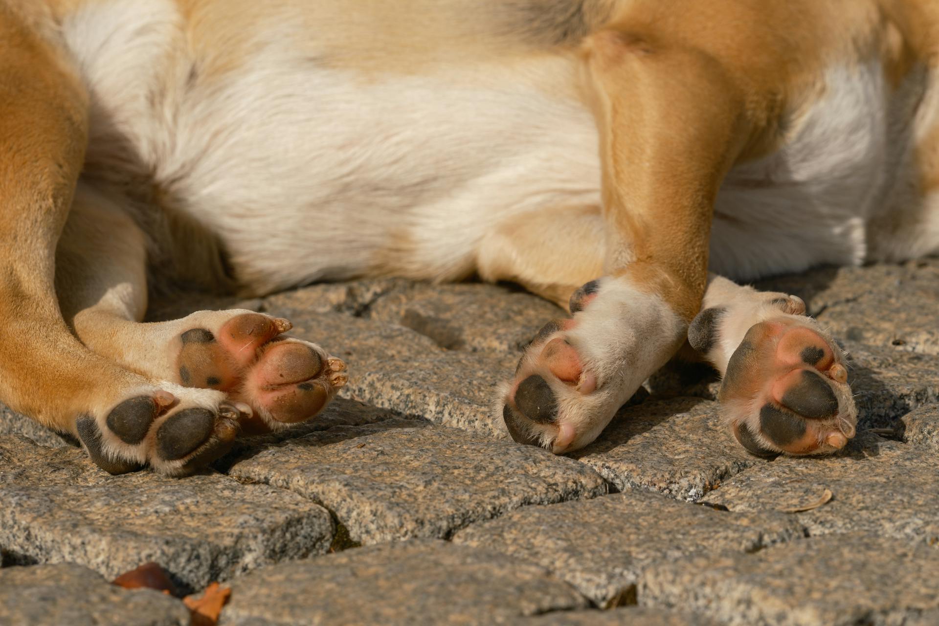
Explore further: Canine Foot Anatomy
The lunate bone is a small, crescent-shaped bone that helps to form the wrist joint. It's located between the scaphoid and triquetrum bones. The lunate bone plays a key role in allowing the dog's front paw to flex and extend.
The triquetrum bone is a small, wedge-shaped bone that helps to form the wrist joint, along with the lunate bone. It's located on the medial (inner) side of the wrist. The triquetrum bone helps to stabilize the wrist joint and allows for movement.
The pisiform bone is a small, pea-shaped bone that's located on the medial (inner) side of the wrist. It's the smallest of the carpal bones and doesn't play a major role in supporting the dog's weight or allowing for movement.
The trapezium bone is a small, four-sided bone that helps to form the wrist joint. It's located on the lateral (outer) side of the wrist. The trapezium bone helps to stabilize the wrist joint and allows for movement.
The trapezoid bone is a small, four-sided bone that's located on the lateral (outer) side of the wrist. It's similar in shape to the trapezium bone and helps to stabilize the wrist joint. The trapezoid bone is smaller than the trapezium bone.
The capitate bone is a large, cube-shaped bone that's located in the center of the wrist. It's the largest of the carpal bones and helps to support the dog's weight and allow for movement.
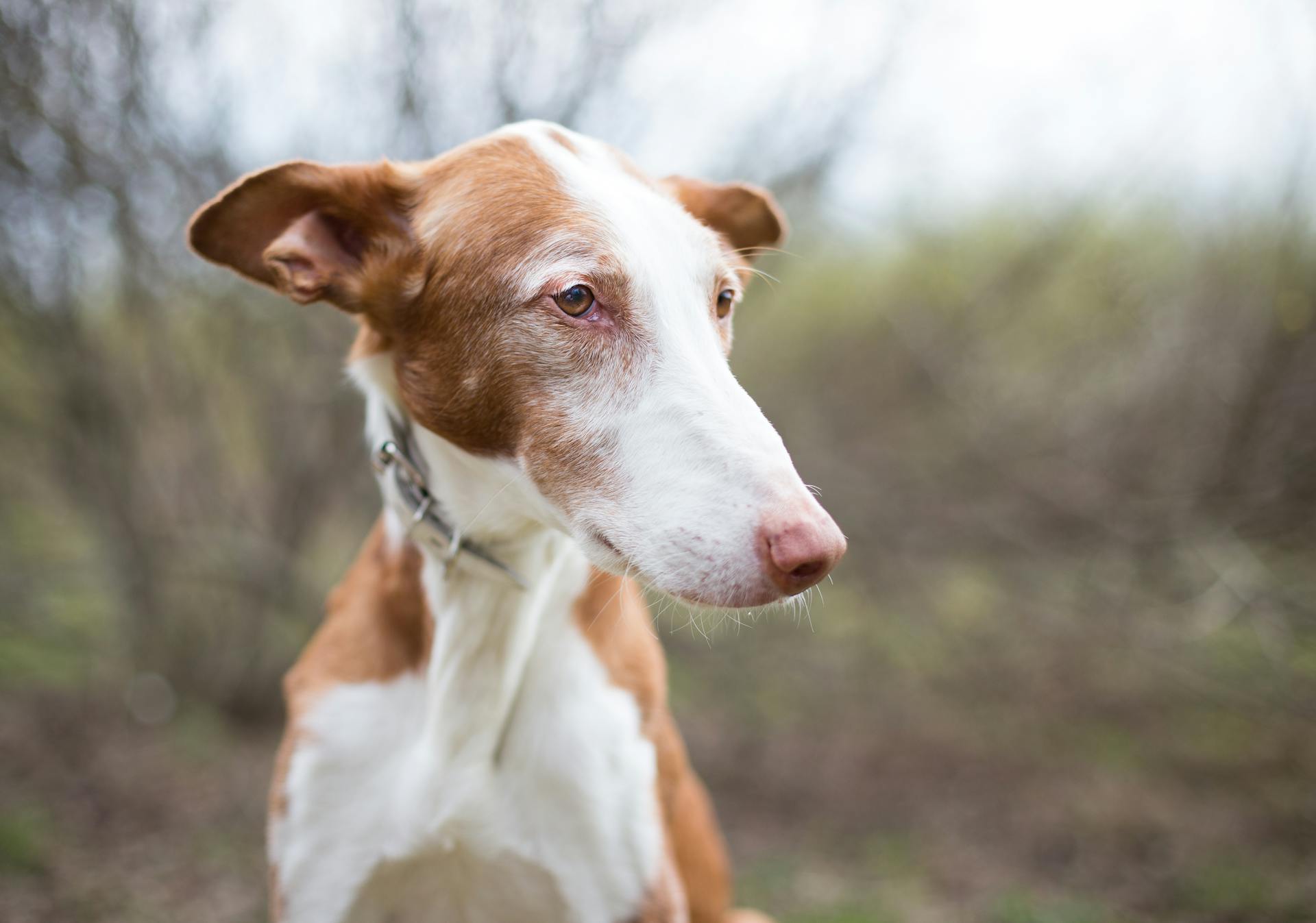
For more insights, see: Why Are Chihuahuas so Small
Carpal Bones
The carpal bones in a dog's front legs are made up of seven bones in total.
Three of these bones are located in the proximal row.
These bones work together to enable a dog's front legs to move in various ways.
The radial and intermediate bones in the proximal row are fused together.
Bones and Joints
The bones in our hands are incredibly complex, with 27 bones in total. The carpal bones, which make up the wrist, are a key part of this structure.
The carpal bones are divided into two rows: proximal (closer to the forearm) and distal (closer to the fingers). This division is crucial for our hand's flexibility and movement.
The proximal row of carpal bones includes the scaphoid, lunate, triquetrum, and pisiform bones. These bones work together to form the wrist's foundation.
The scaphoid bone is the largest bone in the proximal row and is also the most commonly fractured carpal bone. This is often due to a fall onto an outstretched hand.
The distal row of carpal bones includes the trapezium, trapezoid, capitate, and hamate bones. These bones provide a stable base for the metacarpal bones and fingers.
The hamate bone is the shortest bone in the carpal row and is located on the ulnar side of the wrist. It's a vital part of the wrist's structure, allowing for a wide range of motion.
The carpal bones are connected by ligaments, which provide stability and support to the wrist. These ligaments can become stretched or torn, leading to wrist pain and instability.
The scaphoid bone is particularly prone to ligament damage due to its location and function. This can lead to wrist pain and limited mobility.
In summary, the carpal bones are a complex and vital part of our hand's structure. Their unique arrangement and connections enable us to perform a wide range of tasks and movements.
Wrist Bones
The wrist bones are a crucial part of our skeletal system, and they're made up of eight carpal bones.
There are two rows of carpal bones, with four bones in the proximal row and four in the distal row.
The proximal row consists of the scaphoid, lunate, triquetrum, and pisiform bones.
Each of these bones has a unique shape and function, and they work together to form the wrist joint.
The scaphoid bone is the largest of the carpal bones and is located on the thumb side of the wrist.
It's a common injury site, especially for people who fall on their outstretched hand.
The lunate bone is shaped like a crescent moon and is located on the thumb side of the wrist.
It's a relatively small bone, but it plays a big role in wrist movement.
The triquetrum bone is the smallest of the carpal bones and is located on the pinky side of the wrist.
It's a key player in wrist rotation and movement.
The pisiform bone is a small, pea-shaped bone that's also located on the pinky side of the wrist.
It's the only carpal bone that's not connected to the other bones by a ligament.
The distal row consists of the trapezium, trapezoid, capitate, and hamate bones.
Each of these bones has a unique shape and function, and they work together to form the wrist joint.
The trapezium bone is a flat, rectangular bone that's located on the thumb side of the wrist.
It's a key player in thumb movement.
The trapezoid bone is a small, flat bone that's located on the thumb side of the wrist.
It's often overlooked, but it plays a big role in wrist movement.
The capitate bone is the largest of the distal row bones and is located in the center of the wrist.
It's a key player in wrist rotation and movement.
The hamate bone is a small, hook-shaped bone that's located on the pinky side of the wrist.
It's a common injury site, especially for people who play golf or tennis.
Carpal Joint
The carpal joint is a complex structure in a dog's front leg, consisting of three bones: the scaphoid, lunate, and triquetrum. These bones work together to form the wrist joint, allowing for flexibility and movement.
The carpal joint is divided into two parts: the proximal row and the distal row. The proximal row consists of the scaphoid, lunate, and triquetrum bones, while the distal row consists of the radial, intermediate, and ulnar bones.
Functions and Movement
The carpal joint is a complex structure that allows for a wide range of movements in the wrist.
It's made up of eight small bones, known as carpal bones, which are arranged in two rows: proximal and distal.
Each row has four bones, with the proximal row being the ones closer to the forearm and the distal row being the ones closer to the fingers.
The carpal joint is designed to allow for flexion, extension, abduction, adduction, rotation, and circumduction movements.
Flexion is the movement of bending the wrist downwards, like when you're trying to touch your palm to your forearm.
Extension is the opposite movement, where you straighten your wrist outwards.
Abduction is the movement of moving your wrist away from your body, while adduction is moving it towards your body.
Rotation is the movement of rotating your wrist in a circular motion, and circumduction is the movement of moving your wrist in a circular motion, but in a different plane.
The carpal joint is capable of performing these movements because of the unique arrangement of the carpal bones and the surrounding ligaments and tendons.
Joint Structure
Let's take a closer look at the joint structure of the carpal joint.
The carpal bones are arranged in two rows: a proximal row and a distal row.
In the proximal row, there are three carpal bones.
The radial and intermediate carpal bones are fused together.
Joint Function
The carpal joint is a complex structure that relies on proper joint function to move and perform daily tasks smoothly.
The carpal joint is made up of eight small bones in the wrist, which work together to facilitate movement and support the hand.
In a normal carpal joint, the bones fit together like a puzzle, allowing for a wide range of motion.
The joint's function is maintained by a combination of ligaments, tendons, and muscles that surround and support it.
The carpal tunnel, a narrow passageway in the wrist, contains the median nerve and tendons that control finger movement.
Frequently Asked Questions
How do I treat my dogs carpal injury?
For mild carpal injuries in dogs, non-surgical treatment may involve a combination of splints and physical therapy to restore normal function to the affected ligaments. Consult a veterinarian to determine the best course of treatment for your dog's specific condition.
What does carpal hyperextension look like in dogs?
Dogs with carpal hyperextension may display front legs that appear flat-footed or have paws that seem longer than normal
What passes through the carpal canal for dogs?
The carpal canal in dogs contains tendons, nerves, and blood vessels that allow for wrist movement and sensation. Specifically, it houses the flexor tendons, radial and median nerves, and several arteries and veins.
What is carpal arthrodesis in dogs?
Carpal arthrodesis is a surgical procedure that fuses the wrist joint in dogs to alleviate pain and restore function when other treatments have failed. This procedure can significantly improve a dog's quality of life, but it's essential to discuss the risks and benefits with a veterinarian.
Featured Images: pexels.com