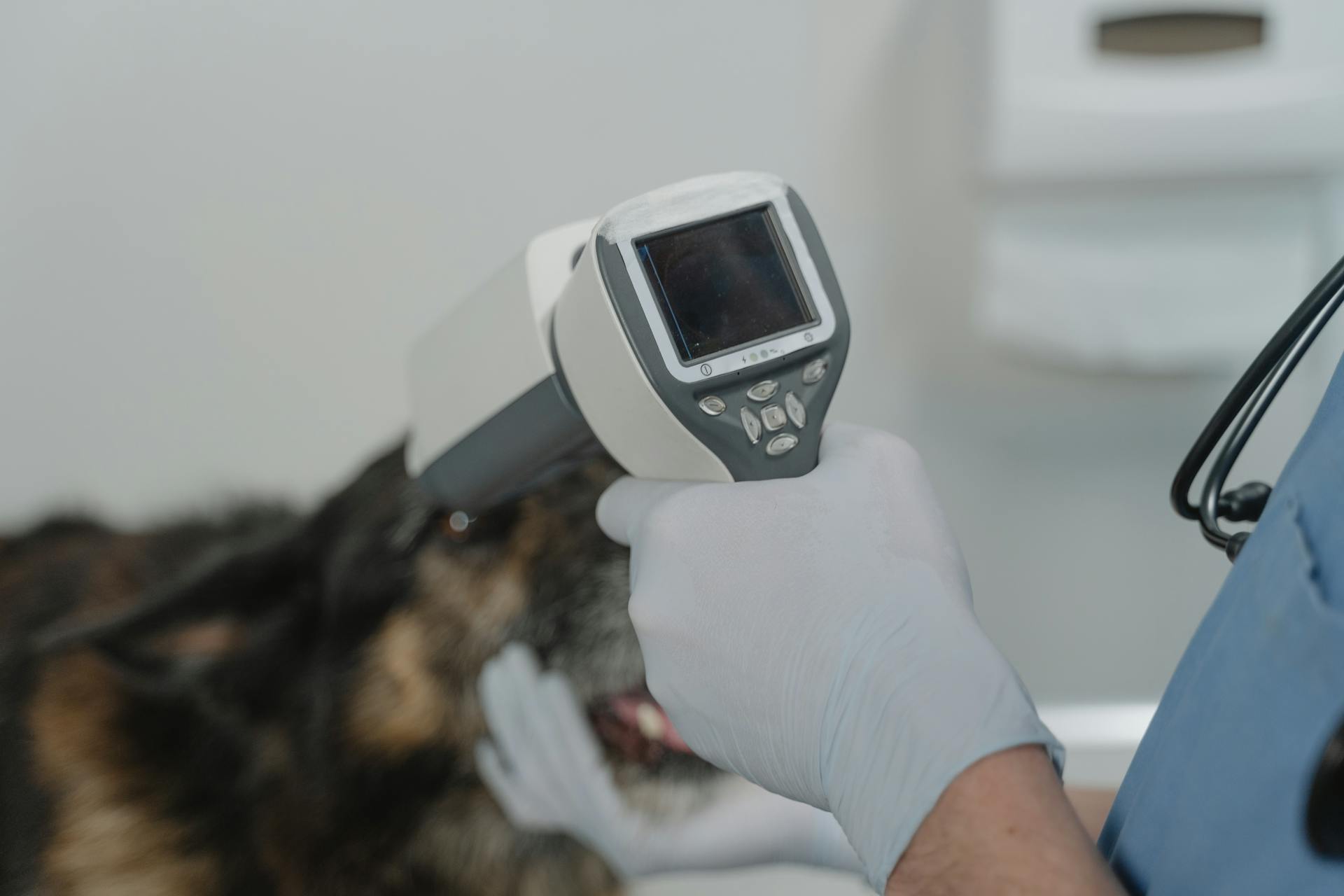
Diagnosing canine liver cancer can be a challenging process, but it's essential to catch the disease early for effective treatment. Liver cancer in dogs can be caused by a variety of factors, including genetics, viruses, and toxins.
Symptoms of liver cancer in dogs may include weight loss, vomiting, and lethargy. These symptoms can be subtle, so regular check-ups with your veterinarian are crucial.
A definitive diagnosis of liver cancer in dogs is typically made through a combination of imaging tests, such as ultrasound and CT scans, as well as a biopsy. The biopsy is a crucial step in confirming the presence of cancerous cells in the liver.
The prognosis for dogs with liver cancer varies depending on the stage and severity of the disease. In some cases, surgery may be possible to remove the tumor, but in many cases, the cancer has spread too far.
Consider reading: Canine Liver Cancer Symptoms
Causes and Risk Factors
Most dogs are diagnosed with liver cancer at the age of 10 or older, making age a significant risk factor.
Expand your knowledge: Pedigree Dog Age Converter
Liver cancer can occur at any age, but it's less common in younger dogs.
Diseases that affect the liver, such as inflammation, liver damage, and Cushing's disease, may increase the chances of developing liver cancer.
Several cancers that can spread to the liver have known breed predispositions, including hemangiosarcoma in Poodles, German Shepherds, Rottweilers, and Golden Retrievers.
Liver cancer in dogs usually affects dogs over 9 years old, with a median age of 11 years for benign liver tumors and 12 years for malignant liver tumors.
Male dogs may be at a higher risk for HCC, although more research is needed to confirm this claim.
Rottweilers are at higher risk of canine lymphoma, which can lead to liver tumors, and female dogs may be at greater risk than males for metastatic liver cancer due to ovarian cancer or mammary gland cancer.
Suggestion: Boston Terrier Brain Tumor Symptoms
Symptoms and Diagnosis
Liver cancer in dogs can be challenging to diagnose, especially in its early stages. In fact, many dogs are asymptomatic until the disease has advanced significantly.
Common symptoms of liver cancer include lethargy, vomiting, diarrhea, fever, increased urination, excessive thirst, loss of appetite and weight loss, jaundice, and weakness. Some dogs may also exhibit non-specific signs such as hyporexia, anorexia, vomiting, weight loss, and lethargy due to the mass effect of a large hepatic tumour.
Diagnostic imaging tests, such as ultrasounds or radiographs, may be used to visualize the liver and detect any abnormalities. Fine needle aspiration or biopsy of the liver may also be necessary to determine if cancerous cells are present. Your veterinarian may also run lab tests, including a urine sample test, to look for signs of liver dysfunction.
Here are some common symptoms of liver cancer in dogs:
- Abdominal pain
- Collapse and acute shock due to tumor rupture
- Decreased appetite
- Diarrhea
- Enlarged or distended abdomen due to fluid accumulation in the abdomen (hemoabdomen)
- Excessive thirst
- Increased urination
- Jaundice or icterus (yellowing of the skin, eyes, and gums)
- Lethargy
- Vomiting
- Weakness, poor coordination, or dullness
- Weight loss
Symptoms of
Symptoms of liver cancer in dogs can be quite subtle, and often don't appear until the disease has advanced. In fact, one in four dogs with liver or bile duct tumors may have no symptoms at all.
Expand your knowledge: Liver Brittany Spaniel

Dogs with liver cancer may exhibit non-specific symptoms that could be caused by many other health problems. These symptoms can include abdominal pain, vomiting, diarrhea, and lethargy. Some dogs may also experience weight loss, decreased appetite, and excessive thirst and urination.
Jaundice, or yellowing of the skin, eyes, and gums, is another common symptom of liver cancer in dogs. This is caused by a buildup of bilirubin in the blood. In some cases, dogs with liver cancer may also experience seizures or a palpable hepatic mass, which is a liver tumor that can be felt during a physical examination.
Here are some common symptoms of liver cancer in dogs:
- Loss of appetite
- Vomiting and/or diarrhea
- Weight loss
- Lethargy and/or weakness
- Increased thirst and/or increased urination
- Ascites (when the abdomen is bloated with fluid)
- Jaundice (yellow tint to the eyes, skin, and gums)
- Seizures
It's worth noting that some dogs may not show any symptoms at all, even if they have liver cancer. In these cases, a veterinarian may detect liver abnormalities during a routine physical exam, such as liver enlargement or abnormal blood work results.
If this caught your attention, see: Liver Mini Schnauzer
Blood Chemistry and CBC
Blood Chemistry and CBC play a crucial role in understanding your dog's health.
Blood cell counts may show a decrease in red blood cell numbers and platelet abnormalities if liver cancer is present.
Blood chemistry will often reveal changes that are a consequence of damage to the liver and gallbladder.
However, the severity of changes in bloodwork does not always correspond to the severity of liver disease in dogs; dogs with liver tumors may have normal blood work or only mild elevations.
For your interest: Mast Cell Tumor in Pit Bulls
Diagnostic Methods
Diagnostic imaging is a crucial tool in diagnosing canine liver cancer, allowing veterinarians to visualize the tumor and assess its size, location, and spread.
Ultrasound is often the first imaging technique used, as it's a painless and non-invasive procedure that provides detailed images of the liver and other internal organs. However, it may not be sufficient to determine the tumor type and whether it's benign or malignant.
Fine needle aspirate (FNA) and cytology are also used to collect a small number of cells from the liver mass, which are then examined under a microscope for cancerous cells. A correct diagnosis is made 60% of the time with liver aspirates.
Diagnostic imaging tests such as ultrasounds or radiographs may also be completed to help veterinarians visualize the tumor and assess its spread.
Suggestion: Lifespan of Dog with Mast Cell Tumor
Bile Acid Testing
Bile Acid Testing is a valuable tool for assessing liver function in dogs. The test measures the bile acid level in a dog's blood before and after eating a fatty meal.
This test helps veterinarians understand if the liver and gallbladder are working normally. If the test is abnormal, it doesn't necessarily mean cancer is the cause, as other liver diseases can also lead to abnormal results.
A fresh viewpoint: Canine Parvovirus Test
Coagulation Profiles
The liver plays a crucial role in producing components for normal blood clotting, so altered liver function can lead to abnormal clotting.
In some cases of liver cancer, the coagulation profile may be normal, but increased prothrombin time (PT), partial thromboplastin time (PTT), and fibrinogen increases are commonly seen.
To prevent abnormal bleeding during procedures like fine needle aspiration, biopsy, or surgery, it's essential to check the coagulation profiles before proceeding.
The liver is very vascular, which is why coagulation profiles are a must-check before any invasive procedures to ensure safe and successful outcomes.
Diagnostic Imaging
Diagnostic Imaging plays a crucial role in diagnosing liver cancer in dogs. Diagnostic imaging is important for tumour localisation, assessment of morphology, staging and surgical planning, and in some cases may allow prediction of histological diagnosis.
Ultrasound is a good way to get a "look" at what's happening in your dog's liver. It's painless and non-invasive, but it does require a specialist with ultrasound training.
Abdominal ultrasound (US) allows confirmation of the presence of a hepatic mass and aids morphological classification. Surgical planning can be performed based on US in smaller well-demarcated tumours but may not be as reliable in larger tumours.
Triphasic CT is often superior to US at assessing liver tumours as it allows detection of smaller lesions and can more accurately assess the relationship between large masses and adjacent soft tissue structures.
MRI and CT scans provide information on the size, location, characteristic features, and extent of tumor spread within the abdomen. These techniques are also helpful in preparing for surgery by allowing the doctor to see the tumor and any involvement of nearby and distant lymph nodes.
Diagnostic imaging tests such as ultrasounds or radiographs may be completed, and a needle aspiration of the liver or biopsy can reveal cancerous cells.
You might enjoy: Boston Terrier Mast Cell Tumor
Types of Liver Cancer
Hepatocellular carcinoma (HCC) is the most common type of liver cancer in dogs, accounting for about 50% of malignant canine liver tumors. It's a slow-growing tumor that can become quite large.
There are different forms of HCC, including massive tumors, nodular tumors, and diffuse tumors. Massive tumors are large, single masses that occur in one liver lobe and have a lower rate of metastasis. Nodular tumors are multiple masses in more than one liver lobe, while diffuse tumors spread throughout the liver lobes.
Here's a breakdown of the different forms of HCC:
Note that diffuse tumors are much harder to treat, while cancers on the left lobe usually have a better chance of successful removal.
Hepatocellular Carcinoma (HCC)
Hepatocellular Carcinoma (HCC) is the most common type of liver cancer in dogs, accounting for about 50% of malignant canine liver tumors.
HCC is a slow-growing tumor that can become quite large, and eventually may cause the liver to bleed within the abdomen.
If left untreated, the tumor may rupture or cause progressive organ function loss, and can compress the liver, leading to end-stage liver disease.
It can also spread to other body areas, such as the local lymph nodes, heart, lungs, or other organs in the abdomen.
Surgery is possible with many HCC tumors, and dogs can recover if the entire mass can be removed, with a better chance of success if the tumor is located on the left lobe.
Large, single masses that happen only in one liver lobe are called “massive tumors” and have a lower rate of metastasis than diffuse or nodular tumors.
Here are the different forms of HCC:
The prognosis for HCC varies depending on the severity of the cancer, with a poorer prognosis for diffuse or nodular tumors that are harder to treat.
A different take: Canine Cancer Prognosis
Hemangiosarcoma (Hsa)
Hemangiosarcoma is a rare and aggressive form of liver cancer that originates from the cells of the inner lining of blood vessels. It's a type of cancer that can occur anywhere in the body where there are blood vessels, but in dogs, it's a significant concern.
Curious to learn more? Check out: Canine Cancer Blood Test
Hemangiosarcoma of the liver is relatively uncommon, making up about 5% of all cases. However, when it does occur, it can spread quickly to other organs like the brain and heart.
The liver is a common site for hemangiosarcoma to metastasize to, even if it's not the primary location of the tumor. This means that even if the cancer starts elsewhere, it can still end up in the liver.
Survival time for dogs with hemangiosarcoma of the liver is limited unless detected early and treated aggressively.
Bile Duct Tumors
Bile duct tumors can be particularly aggressive, with a high rate of local recurrence and metastasis. This makes treatment challenging, even after surgery.
Prognosis is often poor for dogs with bile duct carcinoma, with a typical survival time of less than six months if the liver tumor(s) cannot be removed.
The difficulty of treating bile duct tumors is compounded by the fact that gallbladder cancer is often highly metastatic and difficult to remove.
A unique perspective: Types of Dog Tumors with Pictures
Frequently Asked Questions
How long can a dog with liver cancer live?
Dogs with liver cancer typically live for about two years, with a median survival time of 707 days. However, individual survival times may vary depending on various factors, including the tumor subtype and number of lobes involved.
Is a dog with liver cancer in pain?
A dog with liver cancer may experience abdominal pain due to tumor growth or obstruction of the bile duct. Pain can be a symptom of liver cancer in dogs, but its severity and presence can vary depending on the individual case.
When should I euthanize my dog with liver cancer?
Consider euthanizing your dog if liver cancer causes unmanageable pain, incontinence, or difficulty breathing, and significantly impacts their quality of life. Consult a veterinarian to determine the best course of action for your pet's specific condition.
Sources
- https://www.veterinary-practice.com/article/diagnosis-treatment-canine-hepatobiliary-liver-tumours
- https://www.ahofstatesville.com/services/dogs/dog-cancer
- https://www.animalhospitalofclemmons.com/site/veterinary-pet-care-blog/2020/08/19/liver-cancer-dogs-symptoms-treatment-options
- https://www.akc.org/expert-advice/health/liver-cancer-in-dogs/
- https://www.dogcancer.com/articles/types-of-dog-cancer/liver-cancer-in-dogs/
Featured Images: pexels.com


