The canine spine is made up of 7 cervical vertebrae, 13 thoracic vertebrae, 7 lumbar vertebrae, 3 sacral vertebrae, and a variable number of caudal vertebrae.
These vertebrae are connected by intervertebral discs, which act as shock absorbers and allow for flexibility in the spine.
The cervical vertebrae are the most mobile part of the spine, with a range of motion that allows dogs to twist and turn their heads.
The thoracic vertebrae are connected to the ribs, which protect the heart and lungs.
The lumbar vertebrae are the strongest part of the spine, supporting the dog's body weight and allowing for movement.
The sacral vertebrae are fused together to form a solid bone, providing a stable base for the spine.
See what others are reading: Canine Thoracic Limb Anatomy
Canine Spine Anatomy
The canine spine is a remarkable structure, and understanding its anatomy is crucial for any dog owner or enthusiast.
Located on the dorsal side, or top, of the dog's body, the canine spine is divided into four main regions.
The neck area, called cervical, is one of these regions, and it's just as susceptible to injuries as the human spine.
The mid-back area, called thoracic, is the second region, and it's where many of the dog's vital organs are located.
The lower back, or lumbar, is the third region, and it's a common area for dogs to experience strain or injury.
Dogs are more stoic when it comes to pain, so injury can be harder to pinpoint.
Recommended read: Anatomy of the Canine Eye
Dog Spine
The dog spine is a complex structure that's similar to the human spine. It's divided into four main regions: the neck, mid-back, lower back, and pelvic spine.
The canine spine is made up of a chain of vertebrae, with 7 cervical vertebrae, 13 thoracic vertebrae, 7 lumbar vertebrae, 3 sacral vertebrae, and 20-23 coccygeal vertebrae. These vertebrae have three main functions: protection of the spinal cord, weight bearing and muscle insertion, and movement.
The lumbar area is located in the lower spine and is composed of seven vertebrae protecting the spinal column. It's where the spinal cord changes shape from a tubelike structure to a collection of nerves.
Dogs are more stoic when it comes to pain, so injury to the spine can be harder to pinpoint. However, understanding dog spine anatomy can help you recognize dog spine problems.
Lumbosacral syndrome occurs in the lumbar and sacral region, where a narrowing of the spinal canal causes nerve compression. This can lead to symptoms like difficulty walking, extreme reaction to touch, or complete paralysis.
Dog Vertebrae Numbers
Dogs have 30 vertebrae in their spine.
Each vertebra is connected to the one next to it by a small joint, allowing for movement and held together by ligaments and muscles.
There are seven vertebrae in the neck, 13 in the thoracic area, and seven in the lumbar area.
Dogs have more vertebrae than humans, including an extra one that forms the tail.
Intervertebral discs are a type of spongy cartilage located between vertebrae, cushioning movement and absorbing shock and vibration.
Dogs have 28 intervertebral discs in their spine, one between each vertebra.
The lumbar area, located in the lower spine, is composed of seven vertebrae protecting the spinal column.
A dog's lumbar area connects to the pelvic spine, called sacral.
Comparative Anatomy
The canine spine is made up of 7 cervical, 13 thoracic, 7 lumbar, and 3 sacral vertebrae, which provide flexibility and support for the dog's body.
Each vertebra has a distinct shape and function, with the cervical vertebrae being the longest and most flexible, allowing for a wide range of motion in the neck.
The thoracic vertebrae are shorter and more rigid, providing stability for the ribcage and allowing for deep breathing.
The lumbar vertebrae are the largest and strongest, supporting the dog's weight and facilitating movement.
The sacral vertebrae are fused together to form a solid, triangular structure that provides a stable base for the spine.
The intervertebral discs, located between each vertebra, act as shock absorbers, cushioning the vertebrae and reducing the impact of movement on the spine.
The spinal cord, a continuation of the brain, runs through the vertebral canal and is protected by the vertebrae and intervertebral discs.
The canine spine is designed to be flexible and adaptable, allowing dogs to twist, bend, and move with ease.
Muscle and Disc Anatomy
Let's take a closer look at the muscle and disc anatomy of the canine spine. The spine is supported by 30 muscles, which work together to provide flexibility and stability.
The transverse processes of the vertebrae serve as attachment points for these muscles. The intervertebral discs, on the other hand, act as shock absorbers and allow for flexibility in the spine.
The discs are made up of a tough outer layer called the annulus fibrosus and a softer inner layer called the nucleus pulposus. This unique composition enables the discs to absorb and distribute pressure.
Specimens
The specimens used in this study were six young adult dogs, ranging in weight from 19 to 28 kg, with a mean weight of 23.8 kg.
These dogs were of mixed breed and had a lean to moderate body condition score.
They were estimated to be between 2 and 5 years of age based on their dentition.
All of the animals were healthy, as confirmed by an ante-mortem physical examination.
Muscle Types (Hypaxial & Epaxial)
Muscles in the human body can be broadly classified into two main types: hypaxial and epaxial muscles. These muscle types have distinct origins and insertions, and play different roles in movement and stability.
Hypaxial muscles originate from the anterior (front) part of the vertebral column and insert into the ribs and sternum. They are responsible for movements such as flexion and extension of the vertebral column.
Epaxial muscles, on the other hand, originate from the posterior (back) part of the vertebral column and insert into the transverse processes of the vertebrae. They are involved in movements such as lateral flexion and rotation of the vertebral column.
The hypaxial muscles are further divided into two subgroups: the abdominal muscles and the intercostal muscles. The abdominal muscles include the rectus abdominis, external obliques, and internal obliques, which work together to flex the vertebral column.
Figure 2
Figure 2 shows the anatomy of a spinal disc. The spinal disc is a flat, round structure that acts as a shock absorber between the vertebrae.
The disc has a tough outer layer called the annulus fibrosus, which provides structural support. This layer is composed of strong, fibrous tissue.
The spinal disc also has a soft, gel-like center called the nucleus pulposus. This nucleus is made up of a type of cartilage called hyaline cartilage.
The nucleus pulposus is responsible for absorbing shock and allowing for flexibility in the spine. It's like a big, squishy cushion that helps us move and bend without putting too much pressure on our vertebrae.
The combination of the annulus fibrosus and the nucleus pulposus allows the spinal disc to function properly. If either of these components is damaged, it can cause problems with the spine.
Canine Skeleton
The canine skeleton is divided into two main regions: the axial skeleton and the appendicular skeleton. The axial skeleton consists of the vertebral column and the rib cage.
The vertebral column is a chain comprised of a varying number of vertebrae, with dogs having 7 cervical vertebrae, 13 thoracic vertebrae, 7 lumbar vertebrae, 3 sacral vertebrae, and 20-23 coccygeal vertebrae. This unique arrangement allows for a range of movement and flexibility.
The vertebral column has three main functions: protection of the spinal cord, weight bearing and muscle insertion, and movement.
Canine Skeleton
The canine skeleton is divided into two main regions: the axial skeleton and the appendicular skeleton.
The axial skeleton consists of the vertebral column and the rib cage. The vertebral column is a chain of vertebrae that protects the spinal cord, bears weight, and enables movement.
Dogs have a varying number of vertebrae in their vertebral column, with 7 cervical vertebrae, 13 thoracic vertebrae, 7 lumbar vertebrae, 3 sacral vertebrae, and 20-23 coccygeal vertebrae.
The vertebral column has three main functions: protection of the spinal cord, weight bearing, and muscle insertion. It also enables movement by allowing for flexibility and flexibility.
In total, there are 30 vertebrae in a dog's spine: seven in the neck, 13 in the thoracic, seven in the lumbar, and three in the pelvic area.
Dog Lumbar Location
The dog lumbar location is in the lower spine, composed of seven vertebrae that protect the spinal column. This area is prone to problems, as the spinal cord changes shape here, becoming a collection of nerves.
A dog's lumbar region connects to the pelvic spine, called the sacral region. This connection is a common area for soreness and pain.
Lumbosacral syndrome occurs in this region, caused by a narrowing of the spinal canal, which can lead to nerve compression. This compression can be due to various factors, such as arthritis or a spinal tumor.
Pressure and pain are the results of this compression, and can manifest in a range of symptoms, from difficulty walking to complete paralysis.
Intravertebral Disc Disease
Dogs can experience a slipped disc in the spine, also called intervertebral disc herniation, just like people.
This condition occurs when the intervertebral disc, a cushion between the vertebrae, moves out of place and puts pressure on the spinal cord, causing pain and damage to the nerves.
Bleeding can create additional pressure, making the condition even more serious.
Signs of a slipped or herniated disc in a dog include loss of coordination, weakness or lameness in the back end, incontinence, paralysis, crying, reaction to touch, and personality changes.
A low head and rounding of the back are common clues to spinal issues.
Treatment of IVDD in dogs typically involves oral nonsteroidal anti-inflammatory drugs or corticosteroids, analgesics, muscle relaxants, and/or gabapentin along with activity modification and physical therapy.
Dog Herniated Disc
Dogs can experience a slipped disc in the spine, also called intervertebral disc herniation.
The intervertebral disc is a cushion between the vertebrae offering a padding between the bones in the spine.
Signs of a slipped or herniated disc in a dog include loss of coordination, weakness or lameness in the back end that is often mistaken for leg or knee injury.
Incontinence, paralysis, crying, reaction to touch, and personality changes are also common symptoms.
A low head and rounding of the back are common clues to spinal issues.
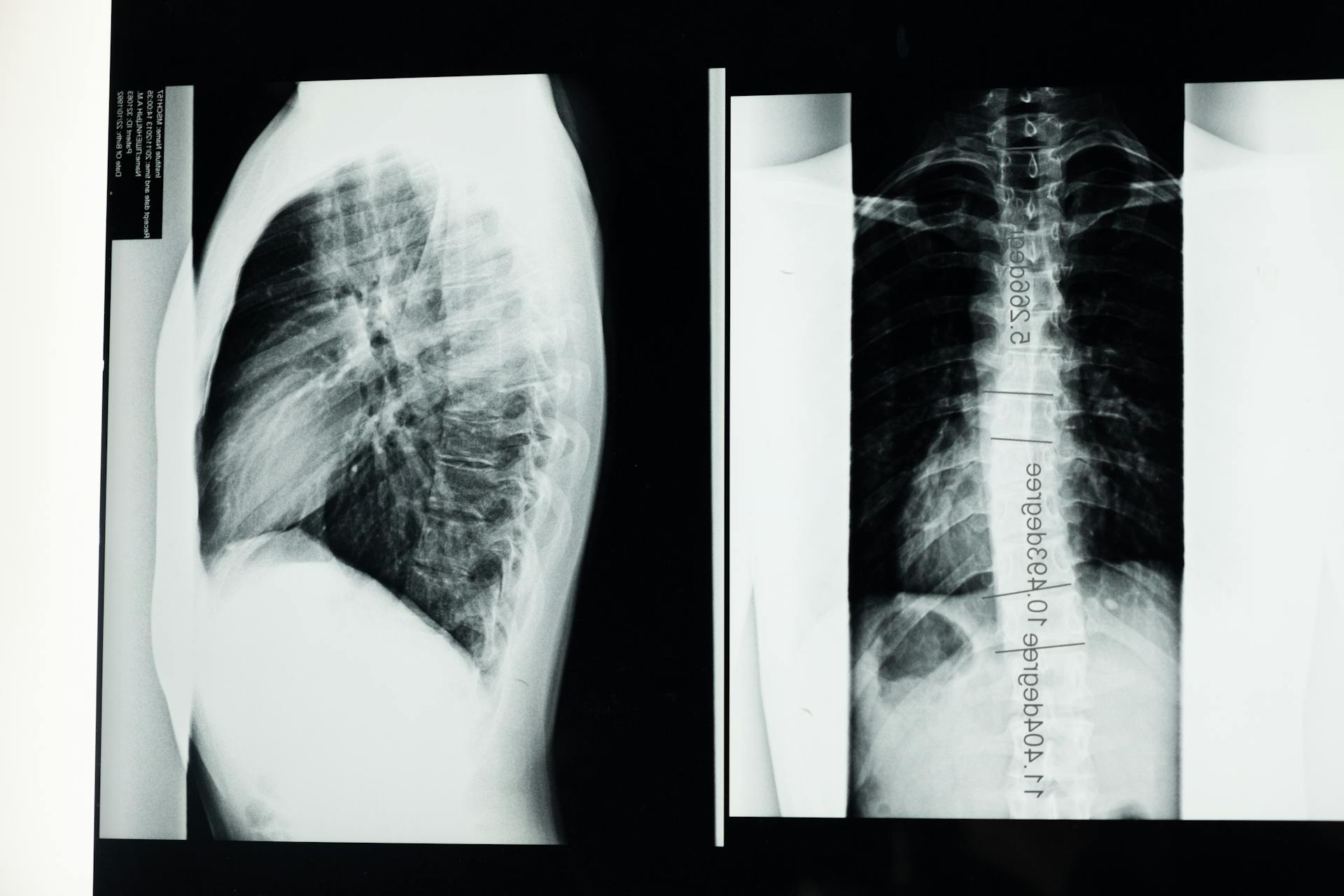
A CT scan or MRI might be necessary to determine the extent of the compression.
Clearly, veterinary attention is required because spinal injuries are serious.
Ivdd in Dogs
Intravertebral Disc Disease (IVDD) in dogs is a serious condition that requires prompt attention. Treatment of IVDD in dogs mimics standard of care therapeutic algorithms for human patients.
Symptoms of pain, stiffness, and muscle spasm without significant neurologic deficits are typically treated with oral nonsteroidal anti-inflammatory drugs or corticosteroids, such as prednisone. Analgesics like tramadol, muscle relaxants, and gabapentin are also commonly used.
Corticosteroid, opioid, and/or local anesthetic epidural, sacroiliac, and facet joint injections have been performed with success in treating IVDD in dogs. Acupuncture and chiropractic treatments have been advocated by some, but evidence for safety and efficacy is currently lacking in veterinary medicine.
Surgical treatment for symptomatic IVDD is indicated when nonsurgical treatment has failed, significant neurologic deficits are present, and/or the pathology necessitates it. The most common indication for surgical treatment of canine IVDD is acute disc extrusion in CD dogs.
Surgical decompression and partial discectomy via partial corpectomy, laminectomy, hemilaminectomy, facetectomy, or foraminotomy of the affected disc space(s) is typically performed to treat acute disc extrusion in CD dogs.
For more insights, see: Dogs Muscle Anatomy
Sources
Featured Images: pexels.com