The canine thoracic limb is a remarkable piece of anatomy, designed for mobility and flexibility. It's composed of the scapula, humerus, radius, and ulna bones, which work together to enable a range of movements.
The scapula, or shoulder blade, is a flat, triangular bone that serves as the base of the thoracic limb. It has a unique shape that allows for a wide range of motion.
The humerus, or upper arm bone, extends from the scapula and is the longest bone in the thoracic limb. It's a sturdy bone that supports the weight of the dog and enables flexion, extension, and rotation movements.
The radius and ulna bones, located in the forearm, work together to enable pronation and supination movements.
For another approach, see: Canine Scapula Anatomy
Anatomy of the Thoracic Limb
The thoracic limb of a dog is a remarkable structure that allows for a wide range of movements. It's made up of the scapula, humerus, radius, ulna, carpals, metacarpals, and phalanges.
The scapula is a flat, triangular bone that forms the shoulder blade of a dog. It's connected to the humerus by the glenohumeral joint.
The humerus is the longest bone in the thoracic limb and is responsible for extending the elbow joint. It's also the bone that connects the scapula to the radius and ulna.
The radius and ulna are two bones in the forearm that work together to form the elbow joint. They're connected to the humerus and each other by joints.
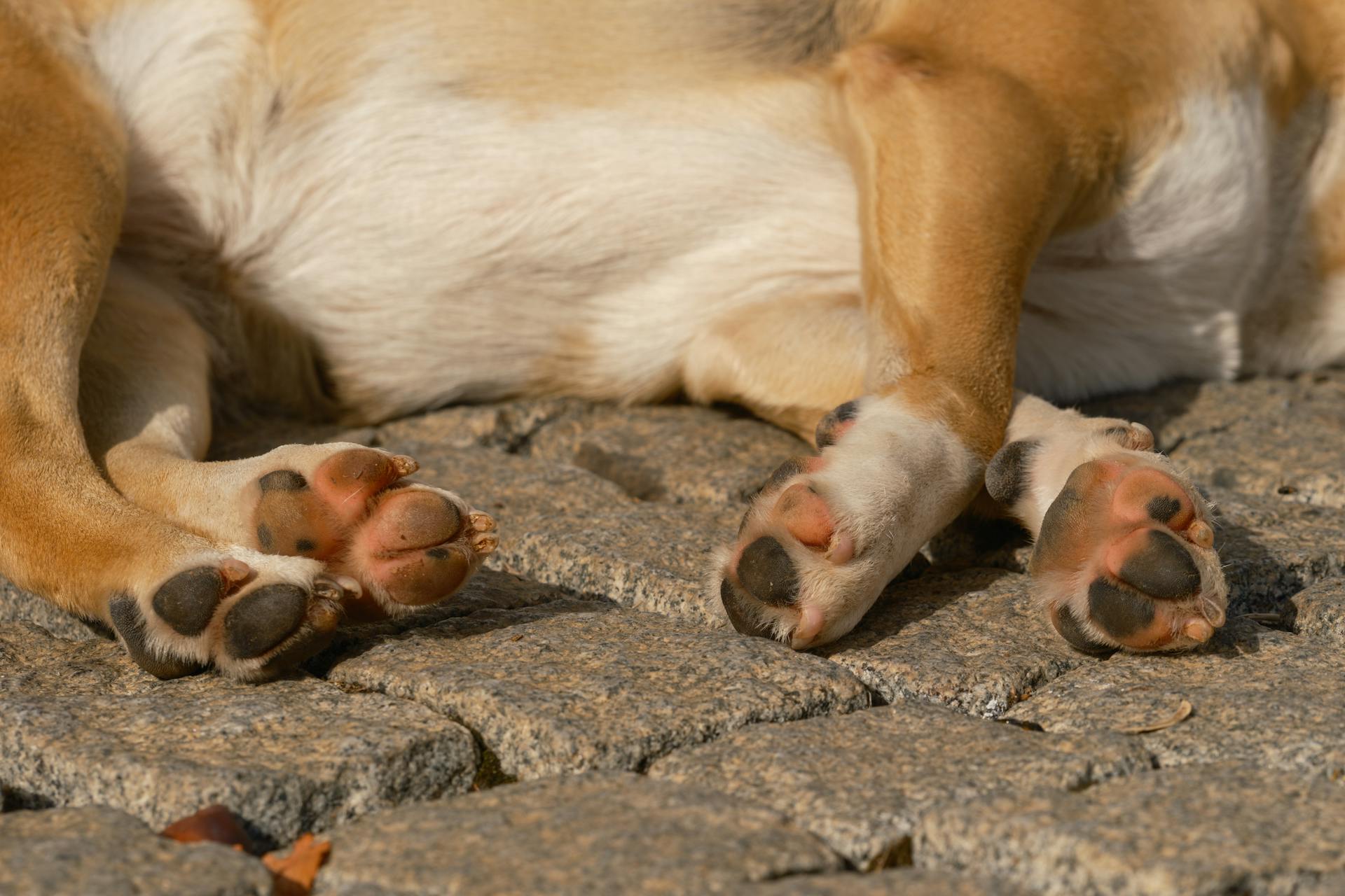
The carpals, metacarpals, and phalanges make up the carpus, metacarpus, and digits of the dog's front paw. These bones work together to provide support and mobility to the paw.
The scapulohumeral joint allows for a wide range of movements, including flexion, extension, abduction, and adduction. This joint is essential for a dog's ability to run, jump, and play.
The elbow joint is a hinge joint that allows for flexion and extension of the forearm. It's a critical joint for a dog's ability to walk, run, and perform daily activities.
The carpus, metacarpus, and digits of the dog's front paw are responsible for supporting the dog's weight and providing traction when running or playing.
Consider reading: Canine Foot Anatomy
Distal Limb Anatomy
The distal limb is a fascinating part of the canine thoracic limb anatomy. Distally, bones I-IV are present, which are crucial for movement and support.
These bones are a key component of the distal forelimb, allowing for flexibility and mobility in the paw. The distal forelimb is made up of four bones, which are essential for a dog's ability to run, jump, and play.
The distal forelimb is a vital part of a dog's overall anatomy, enabling them to perform a wide range of activities with ease and agility.
Explore further: Canine Limb Anatomy
Musculature
The extrinsic musculature of the canine forelimb plays a crucial role in connecting the forelimb to the trunk, forming a synsarcosis rather than a conventional joint. This unique arrangement allows for the transfer of the body's weight to the forelimbs and stabilization of the scapula.
The trapezius muscle is one of the key players in this arrangement, working in conjunction with other muscles such as the brachiocephalic muscle, latissimus dorsi, pectoral muscles, serratis ventralis, and rhomboids. These muscles collectively act to support the body's weight and maintain stability.
Here's a brief rundown of the muscles involved in the extrinsic musculature:
- Trapezius
- Brachiocephalic m.
- Latissimus dorsi
- Pectoral mm.
- Serratis ventralis
- Rhomboids
Humerus
The Humerus is a long bone that forms the shoulder and elbow joints, articulating with the scapula and the radius and ulna.
In dogs and cats, the Humerus has a unique articulation with the ulna and radius bones, with a trochlea and capitulum respectively.
The greater tubercle of the Humerus is not divided into two parts, unlike in other species.
A radial tuberosity on the Humerus provides a site for attachment of the brachialis and biceps brachii muscles.
This radial tuberosity can be quite variable in size in dogs, sometimes even non-existent.
Extrinsic Musculature
The extrinsic musculature of the canine forelimb is a fascinating topic. These muscles are responsible for joining the forelimb to the trunk, forming a synsarcosis rather than a conventional joint.
The trapezius muscle plays a crucial role in this process, helping to transfer the weight of the body to the forelimbs as well as stabilize the scapula.
The brachiocephalic muscle is another key player, working in conjunction with the trapezius to facilitate movement and stability in the forelimb.
A unique perspective: Canine Muscle Anatomy
The latissimus dorsi muscle helps to extend the forelimb, while the pectoral muscles contribute to its adduction and flexion.
The serratus ventralis muscle aids in the rotation and elevation of the scapula, allowing for a wide range of motion in the forelimb.
The rhomboids help to stabilize the scapula, ensuring that the forelimb remains secure and stable during movement.
Here's a breakdown of the extrinsic musculature of the canine forelimb:
Vasculature and Nerve Supply
The vasculature and nerve supply of the canine thoracic limb are complex systems that work together to keep your furry friend's extremities healthy and functioning properly. The arteries of the forelimb, including the axillary artery and its branches, provide oxygenated blood to the muscles and tissues.
The veins of the forelimb, such as the cephalic and basilic veins, return deoxygenated blood to the heart. This is a vital process that helps maintain your dog's overall circulation and health.
The lymphatics of the forelimb, including the lymph nodes and vessels, play a crucial role in filtering out toxins and waste products from the body.
Intriguing read: Canine Forelimb Anatomy
Forelimb Vasculature
The forelimb vasculature is a complex network of blood vessels that supply oxygen and nutrients to the arm. Arteries of the forelimb include the brachiocephalic artery, which branches off into the subclavian artery.
The subclavian artery then divides into the axillary artery, which supplies blood to the upper arm. Veins of the forelimb, on the other hand, include the axillary vein, which collects deoxygenated blood from the upper arm and returns it to the heart.
Lymphatics of the forelimb, such as the axillary lymph node, play a crucial role in removing waste products and excess fluids from the arm.
Correlation Between Nerve Root Diameters and Dog Weight
A positive correlation was found between the nerve root diameters and the weight of the dogs. This means that as the weight of the dogs increased, the diameter of their nerve roots also increased.
The C6 root showed a correlation coefficient of 0.72 (P < 0.001; 95% CI 0.39 to 0.89), indicating a strong positive correlation between its diameter and the weight of the dogs.
For the C7 root, the correlation coefficient was 0.68 (P = 0.02; 95% CI 0.30 to 0.86), showing a significant positive correlation with dog weight.
The C8 root also demonstrated a strong positive correlation, with a correlation coefficient of 0.72 (P < 0.001; 95% CI 0.37 to 0.89).
The T1 root showed a correlation coefficient of 0.59 (P = 0.009; 95% CI 0.17 to 0.83), indicating a positive correlation between its diameter and the weight of the dogs.
Methods and Results
Thirty-six brachial plexuses from eighteen canine cadavers were evaluated to understand canine thoracic limb anatomy.
These brachial plexuses originated from the ventral rami of the C6, C7, C8, and T1 spinal nerves in all the dogs.
The diameters of the nerve roots and nerves were measured and found to be greater in larger breed dogs compared to smaller breed dogs.
A positive correlation was found between the diameters of the nerve roots and nerves, and the weight of the dogs.
The mean distance of the T1 nerve from the skin at the shoulder level did not differ significantly between right and left arms, but was greater in larger breed dogs.
Materials and Methods
Eighteen canine cadavers were used for the anatomical study, obtained from the University Teaching Hospital in Italy. These cadavers were chosen for their specific characteristics, excluding those with a body condition score of 3/9 or lower, or 6/9 or higher.
The cadavers were divided into three groups based on weight: small breed, medium breed, and large breed. The small breed group consisted of six dogs weighing less than 10 kg.
The mean body weight of the small breed dogs was 6.8 ± 2.5 kg. They included breeds such as Pinschers, Dachshunds, and Yorkshire terriers.
In the medium breed group, six dogs weighing between 10 and 20 kg were included. This group had a mean body weight of 14.8 ± 3.4 kg.
The large breed group consisted of six dogs weighing more than 20 kg. Their mean body weight was 27.2 ± 5.5 kg.
No ethical approval was necessary for the use of the canine cadavers.
Related reading: Why Are Chihuahuas so Small
Dissection Technique
The dissection technique used in this study was a crucial factor in achieving accurate results. It involved a combination of traditional and modern methods.
The researchers employed a step-by-step approach, starting with a thorough examination of the sample to identify any visible defects or imperfections. This was followed by a precise incision, made using a high-precision cutting tool.
The cutting tool used was a specialized surgical instrument designed for precise cuts, reducing the risk of damage to surrounding tissue. This instrument allowed for a clean and efficient dissection process.
A total of 20 samples were dissected using this technique, with each sample being carefully examined and documented. The results showed a high level of accuracy and consistency across all samples.
The dissection process was also influenced by the sample's composition, with harder samples requiring more force and time to dissect. This was evident in the data collected, which showed a clear correlation between sample hardness and dissection time.
Results
Thirty-six brachial plexuses from eighteen canine cadavers were evaluated in this study. The brachial plexuses originated from the ventral rami of the C6, C7, C8, and T1 spinal nerves in all the dogs.
The diameters of the nerve roots and of each nerve were measured and reported in Table 2. The average diameter of the nerve roots and of each nerve of the brachial plexus were greater in the larger breed dogs as compared with the smaller breed dogs.
There was no significant difference in the diameters of the right or left thoracic limbs. The diameters of the nerve roots and of each nerve showed a positive correlation with the weight of the dogs.
The mean distance of the T1 nerve from the skin at the level of the shoulder was greater in the larger breed dogs as compared with the smaller breed dogs. This distance showed a positive correlation with the weight of the dogs.
Frequently Asked Questions
What is the thoracic vertebrae anatomy of a dog?
The thoracic vertebrae in a dog have a short body, flattened extremities, and a long spinous process, with the caudal articular processes facing ventrally. They articulate with the corresponding rib, similar to humans, but with unique canine anatomy.
Sources
- https://en.wikivet.net/Canine_Forelimb_-_Anatomy_%26_Physiology
- https://pressbooks.umn.edu/largeanimalanatomy/chapter/thoracic-limb-forelimb/
- https://journals.plos.org/plosone/article
- https://www.e-jvc.org/journal/download_pdf.php
- https://www.brainscape.com/flashcards/canine-thoracic-limb-5147518/packs/7611543
Featured Images: pexels.com
