Cherry eye in dogs is a common condition that affects the tear ducts. It occurs when the gland that produces tears becomes prolapsed, or bulges, out of its normal position.
The prolapsed gland can be red and inflamed, resembling a cherry, hence the name. It's usually a painful and uncomfortable condition for dogs.
Cherry eye can be caused by a weak or underdeveloped tear gland, which can be inherited from a dog's parents. Some breeds are more prone to cherry eye, such as Cocker Spaniels and Boston Terriers.
Symptoms of cherry eye may include excessive tearing, redness, and swelling of the affected eye.
What's Cherry Eye in Dogs?
Cherry eye in dogs is a condition where the gland of the third eyelid, also known as the nictitating membrane, prolapses or falls out of its normal location.
The third eyelid is a thin membrane that moves across the eye diagonally and contains a small T-shaped cartilage. It's mostly pink with a brown or black edge that's visible in almost all dogs.
The third eyelid has a small tear gland, which produces approximately 30% of a dog's tears. The remaining 70% comes from the lacrimal gland, located above the upper eyelid.
A pink or reddish mass in the corner of the eye is often a sign of cherry eye. This mass is the prolapsed gland of the third eyelid.
Cherry eye can affect one or both of a dog's eyes, and any breed can be susceptible to this condition.
Causes and Signs
Cherry eye in dogs can be a pretty noticeable condition. You'll often see a red, raised mass coming from the lower eyelid.
This mass is actually the third eyelid, which is a normal part of a dog's anatomy. The third eyelid gland produces part of the tear film, and if it's not working properly, your dog might develop dry eye symptoms.
Dry eye can cause thick, gooey tears that are often yellow or white. If left untreated, the cornea can become scarred and pigmented, leading to red and inflamed eyes.
Your dog might squint or rub their eyes due to irritation from the cherry eye. Some dogs might even develop red spots around their eyes.
Cherry eye is often associated with brachycephalic or short-nosed breeds, where the eye doesn't sit back far enough in the bony orbit. This can cause the third eyelid gland to become displaced.
Trauma, such as head, neck, or eye injuries, can also lead to cherry eye. In rare cases, a tumor or mass associated with the third eyelid can cause the condition.
Younger dogs, especially those under a year old, are more likely to develop cherry eye. Purebred and brachycephalic dogs are also more prone to the condition than mixed breed dogs with medium-sized skulls.
Discover more: Can Allergies Cause Swollen Lymph Nodes in Dogs
Diagnosis and Treatment
Diagnosing cherry eye is relatively straightforward, often presenting as a pink or reddish mass in the corner of the dog's eye.
The condition tends to occur in dogs under 2 years old, with 75% of cases occurring in dogs less than 1 year old.
Here's an interesting read: Degenerative Myelopathy Old Dog Back Legs Collapsing
A full ophthalmic exam is performed, checking vision and ocular reflexes, measuring pressure in the eye, and using a fluorescent dye to inspect the eye.
The Schirmer tear test measures tear production by applying a strip of paper to the lower eyelid.
The third eyelid gland may prolapse and go back to the normal position several times before staying out permanently, with the longer it stays out, the more likely it will cause damage to the third eyelid cartilage and inflammation of the gland.
Treatment involves surgical repair, but the ophthalmologist may prescribe topical anti-inflammatory medication first to reduce inflammation.
Surgical repair is usually successful, but 10% of cases need repeat surgery, and re-prolapse is most common in animals that have had previous surgery in the area.
Here's an interesting read: English Bulldog Cherry Eye Surgery Cost
Diagnosing Canine Conditions
Diagnosing cherry eye in dogs is relatively straightforward, often starting with the owner noticing a pink or reddish mass in the corner of their dog's eye. This condition tends to occur in dogs under 2 years old, with 75% of cases occurring in dogs less than 1 year old.

A full ophthalmic exam is typically performed by a vet, which includes checking vision and ocular reflexes, measuring pressure in the eye, and using a fluorescent dye to inspect the eye. The Schirmer tear test is also used to measure tear production by applying a strip of paper to the lower eyelid.
The third eyelid gland may prolapse and go back to the normal position several times before staying out permanently, which can cause damage to the third eyelid cartilage and inflammation of the gland. The longer the gland is prolapsed, the more likely this damage and inflammation will occur.
Both cherry eye and cartilage eversion can resemble each other, with cartilage eversion occurring when cartilage in the third eyelid is bent, which can be congenital or acquired. Large breed dogs are more likely to experience both conditions simultaneously.
Treatment
Treatment for cherry eye involves surgical repair, which is almost 100% effective when performed promptly in a younger dog.

Surgery to correct cherry eye involves creating a small pocket with tissue that the gland is housed in, and then suturing it in place with absorbable sutures.
In older dogs, if the gland has been prolapsed for months or years, it can be quite difficult to repair, and it's not very healthy for the gland to remain out.
Some breeds are more prone to cherry eye recurrence, including the American Bulldog, Boxer, and Mastiff, which may require a different technique or a combination of methods in approximately 5% to 10% of cases.
Surgical repair is required to correct this defect, but the ophthalmologist may prescribe topical anti-inflammatory medication for a period before surgery to reduce inflammation first.
The technique most commonly used to repair this condition is the mucosal pocket technique, which requires a general anaesthetic and stitches the area shut with dissolving sutures.
Unfortunately, no method is 100% effective, and 10% of cases need repeat surgery, with re-prolapse most common in animals that have had previous surgery in the area.
Post-operative complications include infection, haemorrhage, re-prolapse, suture irritation of the cornea, and cyst formation, which may take 1-2 weeks to resolve.
Surgery types to remedy cherry eye include anchoring procedures and pocket/envelope procedures, with at least 8 surgical techniques currently existing.
Consider reading: Gastric Dilatation Volvulus Surgery
Veterinary Care
If you notice your dog's cherry eye, contact your veterinary team right away. They'll perform a thorough eye exam using tools like an ophthalmoscope to examine and visualize the eye.
Your veterinarian may perform a series of tests to diagnose the issue, including tonometry to measure eye pressure, a Schirmer tear test to measure tear production, and a fluorescein eye stain to check for corneal scratches or ulcers.
Surgery is usually the recommended treatment for cherry eye, and your veterinarian may refer you to a veterinary ophthalmologist or eye specialist for the procedure. There are two main surgical methods: replacing the glands and suturing them in place, or making an envelope in the conjunctiva and replacing the gland there.
Both surgical methods help preserve the function of the glands, and are preferred over surgical removal unless there's a tumor or the gland has scarring and is no longer functional.
When to See a Veterinary Ophthalmologist
Seeing a veterinary ophthalmologist is a specialized service that's usually recommended after consulting your regular veterinarian. This is because most vets are skilled in performing the necessary procedures, but certain breeds like Cocker Spaniels, Bulldogs, and Great Danes can present unique challenges.
If your vet is not comfortable doing the surgery, it's likely they'll refer you to a veterinary ophthalmologist. This ensures your dog receives the best possible care.
In some cases, your vet may refer you to a specialist if the third eyelid cartilage is bent. This is a specific issue that requires expert attention.
Here are some situations where a vet might refer you to a veterinary ophthalmologist:
- The gland has been prolapsed for months or years without surgery
- The gland is not staying down despite massaging it back into place
- The gland keeps prolapsing after surgery
- The vet is not comfortable doing the surgery
- The third eyelid cartilage is bent
- The vet is less familiar with the breed
Caring for a Dog
If you notice anything abnormal with your dog's eyes, contact your veterinary team immediately. They'll want to start treatment sooner rather than later, especially if there's potential trauma involved.
Your veterinarian will perform a thorough eye exam using tools like an ophthalmoscope to examine and visualize the eye. This will likely include tonometry to measure eye pressure, a Schirmer tear test to measure tear production, and a fluorescein eye stain to check for corneal scratches or ulcers.
To keep your dog comfortable during recovery, you'll want to keep them quiet and calm. This is especially important if they've had surgery to correct a cherry eye.
In most cases, your dog will need to wear an Elizabethan collar (E-collar or "cone") to keep them from rubbing their eyes. Your veterinarian will usually prescribe eye drops to aid in healing.
Your veterinarian might recommend lubricating eye drops to protect the eye and help make up for the lack of tear production before surgery. These will typically need to be used multiple times per day.
If your dog has had an injury to the eye causing a scratch (also called a corneal ulcer), your veterinarian might prescribe an eye antibiotic to help prevent secondary bacterial infections while the cornea heals.
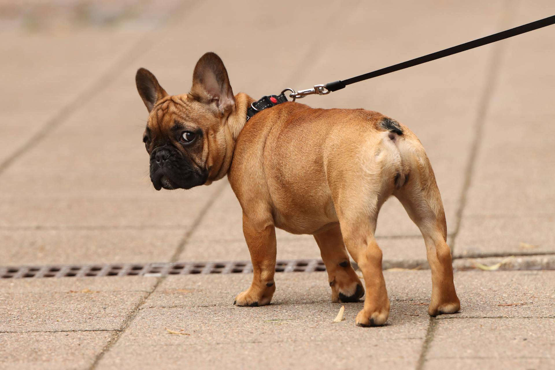
To minimize pressure around the eyes during recovery, it's better to use a harness instead of a collar with an attached leash. This will help keep your dog comfortable and promote healing.
Recommended read: Skin Relief for Dogs with Allergies
Will Repaired Issue Recur?

Some dogs will have cherry eye recurrence, even if the initial surgery was successful.
An estimated 5% to 20% of cases of dogs having surgery will have it recur.
It's essential to discuss the possibility of recurrence with your veterinarian to understand the likelihood of it happening in your dog's case.
The exact percentage of recurrence may vary depending on the individual circumstances of your dog's surgery.
Frequently Asked Questions
Can cherry eye go away on its own?
Cherry eye may resolve on its own with or without medication, but this is not always the case. Surgery is often the most reliable way to treat the condition.
What triggers cherry eye?
Cherry eye in dogs is often caused by weak connective tissue fibers that hold the gland in place, allowing it to slip out and protrude. This condition can be triggered by a combination of genetic and environmental factors.
Sources
- https://www.akc.org/expert-advice/health/cherry-eye-in-dogs/
- https://www.eye-vet.co.uk/veterinary-professional/common-problems/cherry-eye/
- https://en.wikipedia.org/wiki/Cherry_eye
- https://www.dogster.com/ask-the-vet/cherry-eye-in-dogs
- https://www.southernanimalhealth.com.au/veterinary-surgery/cherry-eye/
Featured Images: pexels.com


