ECG Lead Placement is a crucial aspect of cardiac monitoring, and getting it right can make all the difference in diagnosing and treating heart conditions. The ECG Lead Placement Dog, also known as the "ECG Lead Placement Guide", is a tool designed to help healthcare professionals place ECG leads correctly.
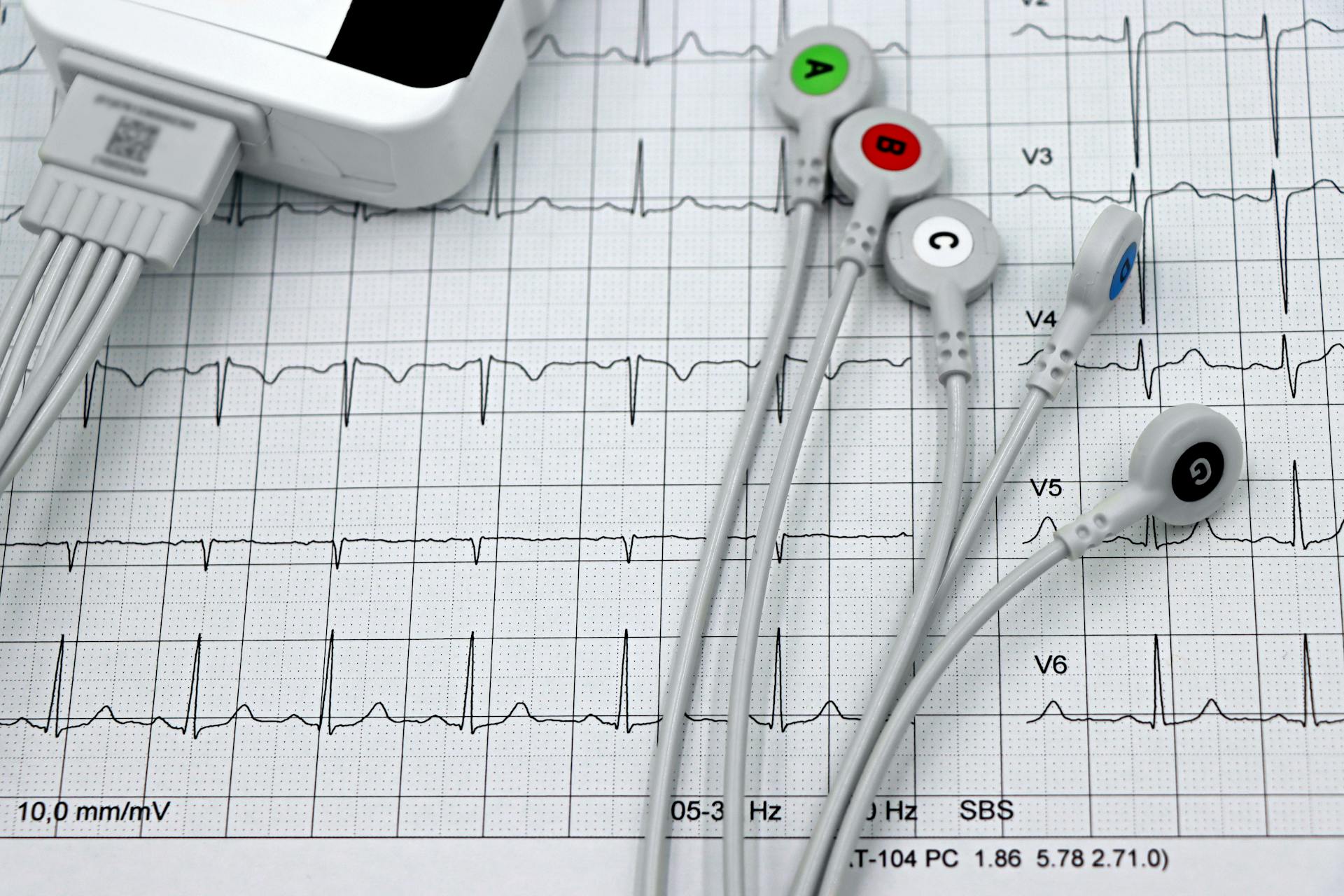
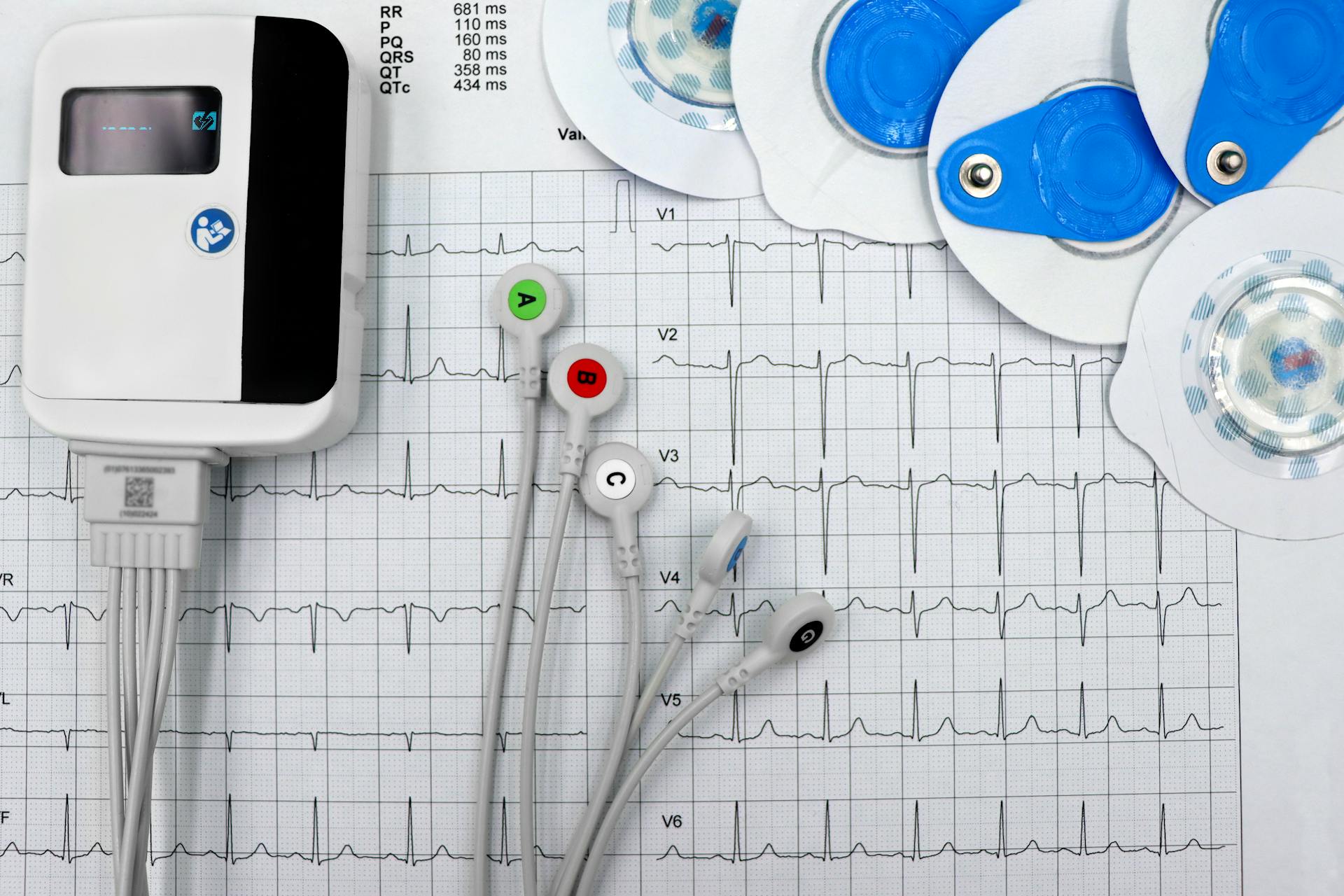
The ECG Lead Placement Dog has six leads, each corresponding to a specific location on the body: I, II, III, aVL, aVR, and aVF. These leads are placed on the patient's chest and limbs to capture the electrical activity of the heart.
A common mistake when placing ECG leads is to confuse the location of the aVR and aVL leads. The aVR lead is placed on the right shoulder, while the aVL lead is placed on the left arm.
Applying ECG Principles to Animals
An electrocardiogram (ECG) is a critical tool in diagnosing and managing heart disease in dogs and cats.
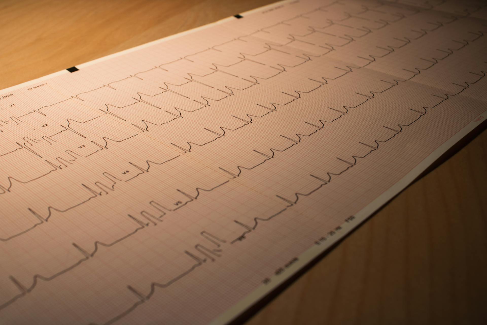
The ECG can be challenging to interpret, even with the best recording, which is why proper lead placement is essential.
To record the electrical potential at different points on the body, ECG leads are placed in specific locations, including the left foreleg, right foreleg, left hindleg, and right hindleg.
For example, Lead 1 places the left foreleg electrode (+) just below the point of the elbow on the back of the left forearm, and the right foreleg electrode (-) just below the point of the elbow on the back of the right forearm.
Lead II places the left hindleg electrode (+) on the loose skin at the left stifle in the region of the patella, and the left foreleg electrode (-) just below the point of the elbow on the back of the left forearm.
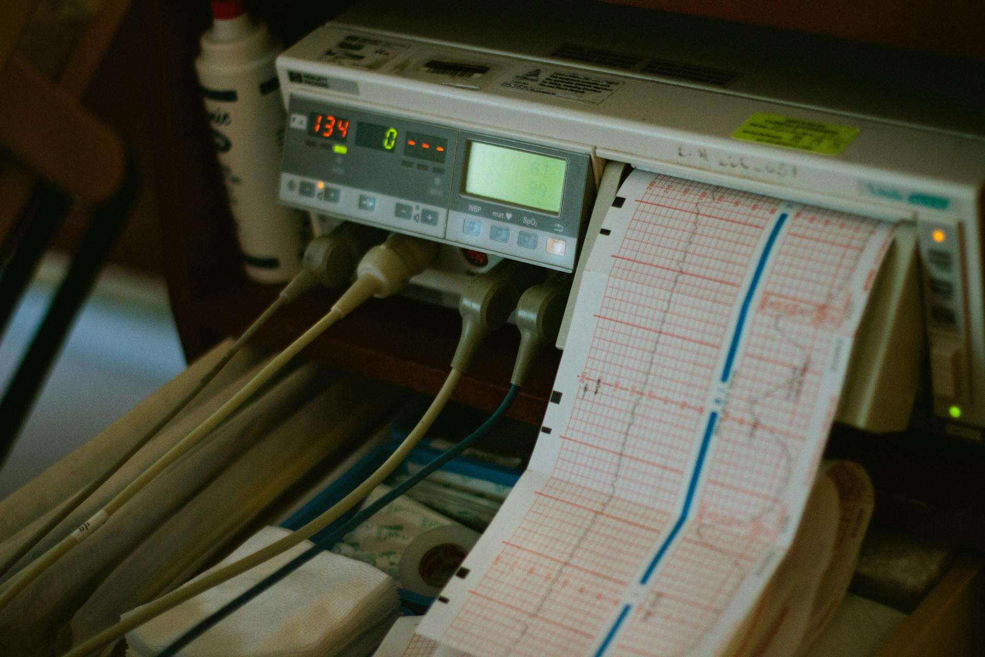
In general practice, there are 3-4 main ECG leads used, although up to 12 leads may be available on a machine.
To remember where to place the leads, you can use the following rhymes: White rhymes with right, so the white lead goes on the right front leg.
Black will go on the other front leg (left), and you read your newspaper in the morning, so the black and white leads will always go on the front legs.
Red goes on the left hind leg, so if black is left front leg, the red is left back leg.
For a Green lead, grass grows on the ground, so the green would go on the right hind leg which is in the down position.
For your interest: Dog Food for White Dogs
ECG Lead Placement on Animals
To place ECG leads on animals, it's essential to follow a specific pattern. ECG leads record the electrical potential at different points on the body, and the placement of leads determines the pattern we're looking for on the recording.
In general practice, we use 3-4 main ECG leads, and we often use an ECG during anesthesia monitoring or when working up cardiac cases. You can remember where to place the leads by using the following rhymes: White rhymes with right, so the white lead goes on the right front leg.
The right forelimb is down in the right lateral position, and snow is on the ground, which helps you place the white lead. You can also use the rhyme "black will go on the other front leg" to place the black lead on the left front leg.
The red lead goes on the left hind leg, and the green lead goes on the right hind leg. You can use the rhymes "smoke over fire" and "grass grows on the ground" to help you remember the placement of the red and green leads.
If this caught your attention, see: Slip Lead Dog Leash How to Use
Key Placement Considerations
When placing ECG leads on animals, it's essential to consider their unique physiology and anatomy.
The size and shape of the animal's chest cavity should be taken into account to ensure proper lead placement.
In dogs, the chest cavity is more shallow than in humans, requiring a shorter lead length.
In cats, the chest cavity is more narrow, making it crucial to position leads carefully to avoid interference.
The location of the animal's heart should be identified before placing leads, typically at the level of the 5th intercostal space in dogs and cats.
A lead placed on the left side of the chest, near the heart, is ideal for obtaining a clear ECG signal.
The position of the lead on the left side of the chest is also important for dogs with a deep chest cavity, such as Great Danes.
In addition to the location, the direction of the lead is also crucial, with leads placed perpendicular to the chest wall for optimal signal quality.
For animals with a more muscular build, such as horses, the lead placement should be adjusted to account for the muscle mass.
In horses, the ECG leads are typically placed on the left side of the chest, between the 5th and 6th intercostal spaces.
The use of multiple leads is often necessary for accurate ECG readings, especially in animals with irregular heart rhythms.
Multiple leads can be placed on the chest and limbs to capture a more comprehensive view of the animal's cardiac activity.
Common Placement Mistakes to Avoid
In general practice, there are 3-4 main ECG leads we use, and it's easy to get them mixed up.
Don't place the white lead on the left front leg, as it's actually supposed to be on the right front leg, where the right forelimb is down.
The black lead should go on the right front leg, not the left, unless you're reading your newspaper in the morning.
Avoid placing the red lead on the right hind leg, as it's actually supposed to be on the left hind leg, where the red and green leads go.
Frequently Asked Questions
How to restrain a dog for an ECG?
To restrain a dog for an ECG, gently hold their forelimbs perpendicular to their body and their hindlimbs in a semiflexed position. This method allows the dog to remain fully conscious without chemical restraint.
Sources
- https://www.sinokmed.com/blogs/news/taking-about-veterinary-ecg-leads-blood-pressures-and-sp02-sensor-placement
- https://blog.vettechprep.com/remembering-where-to-place-ecg-leads
- https://www.theveterinarynurse.com/content/practical/how-to-use-an-ecg-machine/
- https://www.dvm360.com/view/basics-electrocardiography-proceedings
- https://www.cardiobird.com/the-ecg-leads-introduction-and-applications/
Featured Images: pexels.com