Understanding canine hindlimb anatomy is crucial for any dog owner or enthusiast. The hindlimb is made up of the femur, patella, tibia, fibula, and metatarsals.
The femur, or thigh bone, is the longest bone in the hindlimb, extending from the hip joint to the knee. It plays a vital role in supporting the dog's body weight and facilitating movement.
The patella, or kneecap, is a small, triangular bone that protects the joint between the femur and tibia. It's a fascinating example of evolutionary adaptation, as it allows dogs to jump and run with ease.
The tibia and fibula work together to form the lower leg, with the tibia bearing the majority of the body's weight. This is especially important for dogs that are prone to joint issues.
Pelvic Limb Anatomy
The pelvis is a complex bony structure that serves as the foundation for the canine hindlimb, providing stability and support for its movement.
The pelvis is formed by three fused bones: the ilium, ischium, and pubis. These bones work together to form a strong and stable structure.
The ilium, the largest and most dorsal bone, articulates with the sacrum, a triangular bone located at the base of the vertebral column. This connection forms the sacroiliac joint, a strong and stable joint that transmits forces from the spine to the hindlimb.
The ischium and pubis meet to form the acetabulum, a cup-shaped socket that houses the head of the femur, the thigh bone. This joint, known as the hip joint, allows for a wide range of motion.
The pelvis acts as an anchor point for muscles and ligaments, providing stability to the hindlimb during weight-bearing and movement. Its structure and articulation with the sacrum and femur make it essential for supporting the weight of the body.
For another approach, see: Canine Femur Anatomy
Gait and Movement
When evaluating a dog's gait, it's essential to observe for evidence of lameness. Observe the dog for evidence of lameness.
A trained handler should walk and trot the dog back and forth and from side to side on a long, nonslip, distraction-free walkway to confirm the affected limb(s). This is a crucial step in evaluating the dog's gait and movement.
Lameness can be a clear indication of a problem with the canine hindlimb anatomy.
Limb Appearance and Pain
Dogs with pelvic limb lameness may place the lame limb eccentrically, meaning it's farther from midline. This can be a sign of pain or discomfort.
Visible muscle atrophy of the thigh and gluteal muscles can also be a sign of pelvic limb lameness. This can make the affected limb appear smaller compared to the other side.
During a standing evaluation, you can assess for joint effusion, which is swelling in the joint, and palpate the limbs to check for abnormalities.
Consider reading: Canine Limb Anatomy
Evaluate Limb Appearance
To evaluate limb appearance, start by observing how your dog stands naturally. They may place the lame limb eccentrically, or farther from midline, and have visible muscle atrophy of the thigh and gluteal muscles.
Recommended read: Canine Thoracic Limb Anatomy
Evaluate each joint for its standing angle and position, as well as the overall limb for visible deformities and obvious length discrepancies. You can use a measuring tape to provide more objective measurement of muscle mass.
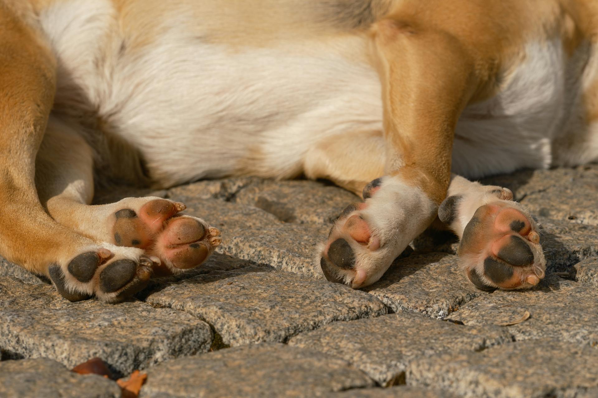
Dogs with pelvic limb lameness often have more of the metatarsal pad visible compared with the contralateral limb. This can be a sign of muscle atrophy.
Subjective assessment of muscle symmetry can be performed by using hands to encircle the thigh and comparing the size of the pelvic limbs. This can help you identify any asymmetries.
Visible deformities and length discrepancies can be a sign of underlying orthopedic conditions. It's essential to note these signs during your evaluation.
Discover more: Canine Anatomy Muscles
Is My Dog's Rear End Elevated?
If your dog's rear end is elevated, the shoulder, thoracic spine, and stifle could be at risk.
This is because a "high in the rear" dog can put uneven pressure on their joints, potentially leading to discomfort or pain.
You might enjoy: Breeds of Dogs with Rear Dew Claws
Some dogs may naturally have a more upright posture, while others may develop it due to muscle imbalances or other underlying issues.
Conditioning exercises can help alleviate this issue by strengthening the muscles in the rear end and improving overall flexibility.
These exercises can also help redistribute the dog's weight more evenly, reducing pressure on sensitive joints.
For example, exercises that target the gluteal muscles can help lower the rear end and promote a more neutral posture.
Specific Joints and Bones
The ilium is a large and prominent bone in canines, with two prominences on the tuber coxae: the cranial and caudal ventral iliac spines. These prominences are readily palpable.
The canine tarsus, or ankle joint, is a complex structure that plays a vital role in hindlimb flexibility. It's a fusion of seven tarsal bones, including the talus, calcaneus, navicular, cuboid, lateral cuneiform, intermediate cuneiform, and medial cuneiform.
The fibula is a slender yet resilient bone that assists the tibia in weight-bearing and stabilizing the ankle. Its unique shape allows it to interlock with the tibia and tarsal bones, forming a sturdy framework that prevents excessive movement and ensures joint stability.
The femur is the longest bone in the hindlimb, forming the vital link between the pelvis and the lower leg. Its connection to the stifle joint, where it meets the tibia and fibula bones, is a testament to its profound impact on mobility.
A unique perspective: Canine Tarsus Anatomy
Hock
The hock is a crucial joint in a dog's hindlimb, responsible for providing flexibility and stability.
It's formed by the tarsus, a complex structure consisting of seven tarsal bones, including the talus, calcaneus, navicular, cuboid, lateral cuneiform, intermediate cuneiform, and medial cuneiform.
These bones are held together by strong ligaments, tendons, and muscles to form a robust joint that allows for a wide range of motion, including dorsiflexion and plantar flexion.
The hock bears the weight of the body during standing and locomotion, and its bones are strong enough to withstand the impact forces associated with these activities.
The tibia forms the main support for the hock, while the fibula provides additional stability.
Palpating the hock is an important part of a veterinary examination, where a veterinarian will apply valgus and varus forces to assess for lateral and medial instability.
The common calcanean tendon inserts on the calcaneus, and palpating it can help identify any abnormalities.
Tibia: Lower Leg Bone
The tibia is a sturdy pillar in the canine hindlimb, extending from the knee joint to the ankle joint and serving as a central axis for the lower leg.
Its cylindrical and slightly curved shaft provides structural support and reinforcement, with a wider proximal end that articulates with the femur and a narrower distal end that connects to the tarsus.
The tibia forms a hinge joint with the femur and a pivot joint with the patella, enabling flexion and extension of the stifle joint.
At its distal end, the tibia articulates with the fibula and the tarsus, contributing to the stability and range of motion of the ankle joint.
The tibia provides attachment points for several muscles, including the gastrocnemius and the tibialis cranialis.
The nutrient artery supplies blood to the bone's tissues through an opening called the nutrient foramen.
Palpating the tibia medially is easiest because soft tissue coverage in this area is minimal.
Palpating the tibia for swelling, pain, or instability is an important step in assessing joint health.
Joints and Synovial Tissues
The stifle joint is a vital part of the canine hindlimb, acting as a hinge to allow for smooth and controlled bending of the hind leg.
Careful localization of pain to the stifle is important, as certain bone tumors are common in this location.
Palpating the stifle for abnormalities, such as CREPI, and evaluating for effusion around the patellar ligament can help identify potential issues.
Assessing for collateral ligament instability by applying varus and valgus force to the stifle with the joint in full extension is crucial for diagnosing early cranial cruciate ligament disease.
The femur, or thigh bone, plays a pivotal role in the dog's ability to navigate its world, forming the vital link between the pelvis and the lower leg.
The femur's connection to the stifle joint is a testament to its profound impact on mobility, ensuring that the joint moves seamlessly and enabling effortless movement.
The tarsus, or ankle joint, is a complex and crucial structure that plays a vital role in a canine's mobility and overall well-being.
See what others are reading: Canine Front Leg Anatomy
The tarsus is situated where the tibia and fibula meet the metatarsus, and consists of seven tarsal bones held together by strong ligaments, tendons, and muscles.
The primary function of the canine tarsus is to provide flexibility and stability to the hindlimb, acting as a hinge joint that allows for a wide range of motion.
The fibula, a slender yet resilient bone, provides unwavering support to the hindlimb's symphony of motion, assisting the tibia in weight-bearing and stabilizing the ankle.
The fibula's distal end contributes to the formation of the lateral malleolus, a bony projection that helps guide the tendons that control foot movement.
The stifle joint, tarsus, and fibula all work together to provide strength, stability, and flexibility to the canine hindlimb, enabling a wide range of motion and essential for everyday activities.
For another approach, see: Norwegian Lundehund Flexibility
Femur
The femur, also known as the thigh bone, is a vital part of a dog's hindlimb anatomy.
It's the longest bone in the hindlimb, spanning from the hip to the knee, and plays a crucial role in mobility.
The femur's connection to the stifle joint, where it meets the tibia and fibula bones, is a testament to its profound impact on movement.
The femur ensures that the joint moves seamlessly, enabling effortless movement, in conjunction with the patella, or kneecap.
The femur forms a harmonious triad with the tibia and fibula, supporting the weight of the dog's body and providing stability for swift and balanced locomotion.
To assess the femur, palpate it for evidence of swelling, pain, or instability, as it's easiest on the lateral aspect between muscle bellies.
The femur's robust structure makes it an essential part of the hindlimb, allowing for a wide range of motions essential for everyday activities.
Bone Specifics
The ilium is a large and prominent bone in canines, with two prominences on the tuber coxae: the cranial and caudal ventral iliac spines.
In dogs, the sacral tuber has two prominences as well: the cranial and caudal dorsal iliac spines.

The iliac crest is wide and convex in canines, and the ileal wing is orientated in an almost sagittal manner.
The femoral head of the canine femur is circular and situated in the centre of the head.
A distinct neck connects the femoral head to the shaft of the femur.
The canine ischial tuberosity is linear in shape and located within the ischium.
The femoral head is level with the greater trochanter in canines.
Femoral palpation is easiest on the lateral aspect between muscle bellies.
The fibula is a slender yet resilient bone in the canine hindlimb, providing support to the tibia and stabilizing the ankle joint.
The fibula's distal end contributes to the formation of the lateral malleolus, a bony projection that guides the tendons controlling foot movement.
Patellar Luxation Assessment
Patellar Luxation Assessment is a crucial evaluation to determine if a joint is functioning properly. To assess for patellar luxation, you'll need to examine the stifle joint.
Medial patellar luxation can be evaluated by extending the stifle, internally rotating the foot, and applying medially directed pressure to the patella. This can help identify any issues with the joint.
Lateral patellar luxation is assessed by partially flexing the stifle, externally rotating the foot, and applying laterally directed pressure to the patella. This will help you determine if the patella is luxated laterally.
Discover more: Canine Stifle Joint Anatomy
The Fibula: A Resilient Support
The fibula is a slender yet resilient bone that plays a crucial role in the ankle joint, known as the tarsus.
It assists the tibia in weight-bearing and stabilizing the ankle, making it a vital component of the canine hindlimb.
The fibula's unique shape allows it to interlock with the tibia and the tarsal bones, forming a sturdy framework that prevents excessive movement and ensures joint stability.
This intricate interplay is essential for the dog's ability to walk, run, and jump with ease.
Here's an interesting read: Canine Tibia Anatomy
The fibula's distal end contributes to the formation of the lateral malleolus, a bony projection that helps guide the tendons that control foot movement.
This complex structure ensures that the ankle joint functions smoothly, allowing the dog to navigate uneven terrain and change direction with agility.
In essence, the fibula provides strength, stability, and flexibility to the canine hindlimb.
Musculature and Support
The pelvis is the foundation for the canine hindlimb, providing stability and support for its movement. It's formed by three fused bones: the ilium, ischium, and pubis.
The ilium, the largest and most dorsal bone, articulates with the sacrum, a triangular bone located at the base of the vertebral column. This connection forms the sacroiliac joint, a strong and stable joint that transmits forces from the spine to the hindlimb.
The acetabulum, a cup-shaped socket, houses the head of the femur, the thigh bone. This joint, known as the hip joint, allows for a wide range of motion.
The pelvis acts as an anchor point for muscles and ligaments, providing stability to the hindlimb during weight-bearing and movement. Its structure and articulation with the sacrum and femur make it essential for supporting the weight of the body.
The proximal aspect of the femur is closer to the pelvis, which is important for understanding the musculature and support of the hindlimb.
Frequently Asked Questions
What are the major nerves of the hindlimb?
The major nerves of the hindlimb include the femoral, obturator, and sciatic nerves, with the sciatic nerve further branching into the tibial and common fibular nerves. Understanding these nerves is crucial for grasping the complex anatomy of the hindlimb.
Sources
- https://www.cliniciansbrief.com/article/pelvic-limb-lameness-canine-dog-bone-joint-orthopedic-exam
- https://en.wikivet.net/Canine_Hindlimb_-_Anatomy_&_Physiology
- https://sciencespace.blog/canine-hindlimb-anatomy/
- https://canineconditioningcoach.com/canine-anatomy-terms/
- https://anatomywarehouse.com/axis-scientific-canine-hindlimb-with-foot-a-109194
Featured Images: pexels.com


