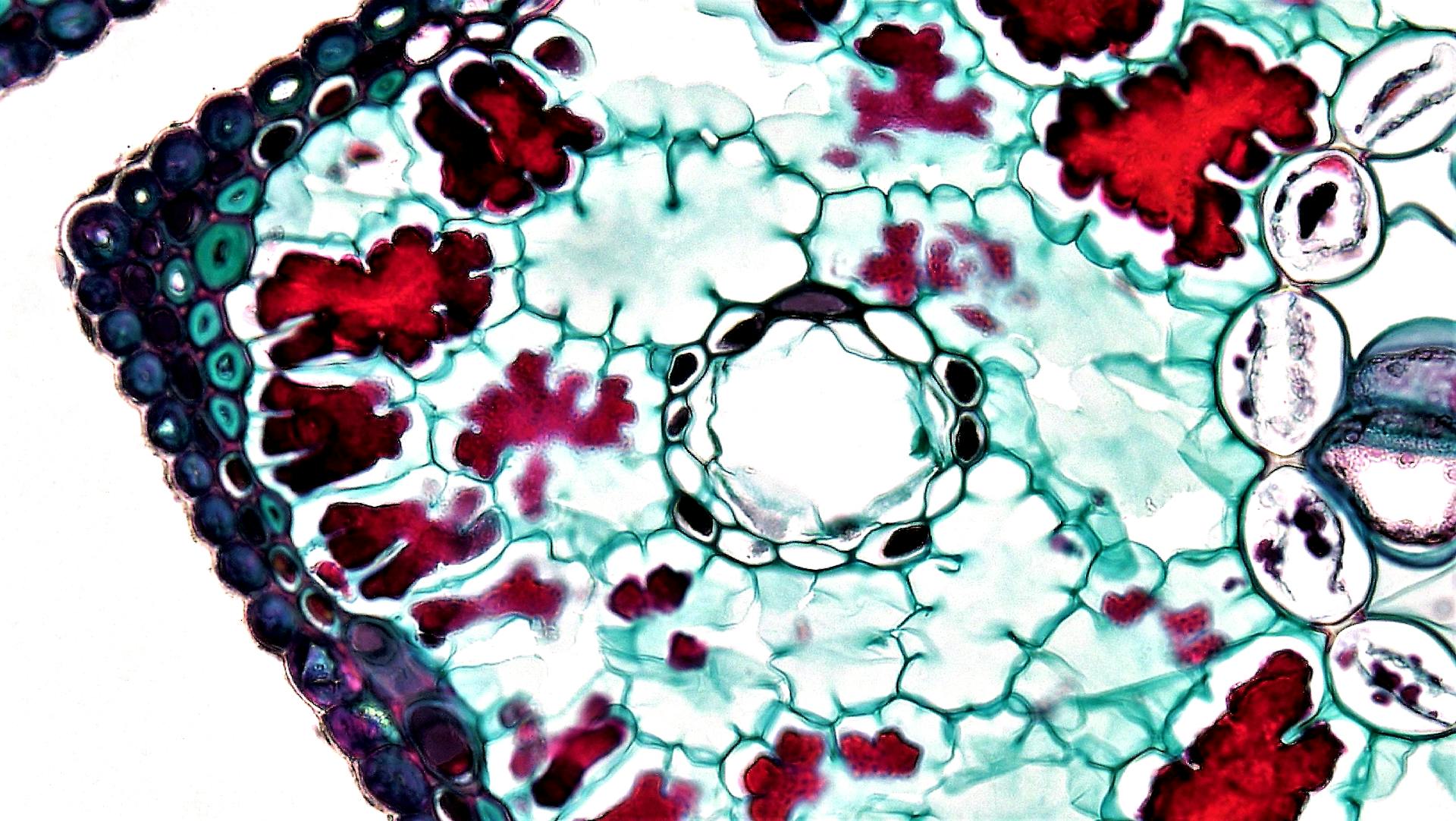
The canine mammary gland is a vital part of a dog's anatomy, responsible for producing milk to nourish their puppies. It's made up of ducts, glands, and fatty tissue.
The mammary gland is divided into four quadrants, with the pectoral muscle forming the boundary between the front and back quadrants. Each quadrant contains 2-5 teats, depending on the dog's size and breed.
The mammary gland is made up of two types of tissue: glandular tissue, which produces milk, and fatty tissue, which provides insulation and support. The glandular tissue is composed of cells that secrete milk, while the fatty tissue helps to protect the gland and provide energy.
The mammary gland's blood supply is crucial for milk production, and it's provided by the internal thoracic artery and vein. This blood supply also helps to regulate the gland's temperature and function.
A different take: Lump on Female Dog Nipple
Canine Mammary Gland Anatomy
The canine mammary gland anatomy is a fascinating topic. The mammary gland of the canine is considered an accessory organ or gland, developing with the influence of hormones during puberty and becoming functioning at the later stage of pregnancy.
Broaden your view: Canine Salivary Gland Cancer
These glands become more developed during late pregnancy and after parturition, enlarging through the action of estrogen and progesterone. Fat deposits, stromal development, and growth of the lobule, ducts, and alveoli are all caused by these hormones.
The anatomical fact of the dog mammary glands divides into two main components: epithelial glandular tissue, along with the connective tissue stroma, and the papillary duct system of the dog's mammary glands.
Here's a breakdown of the main components of the canine mammary gland anatomy:
- Epithelial glandular tissue
- Connective tissue stroma
- Papillary duct system
The papillary duct system is a crucial part of the canine mammary gland anatomy, and it's interesting to note that the lymphatic channels anastomose and encircle the base of the papillary part of the dog mammary glands.
Intriguing read: Mammary Gland Tumor
Anatomical Structure
The canine mammary gland is a complex organ, but understanding its basic structure is key to grasping its function. It's located in the chest area, just beneath the skin.
The mammary gland is made up of ducts, alveoli, and stroma. The ducts are the tubes that carry milk from the alveoli to the nipple, while the alveoli are the tiny sacs where milk production occurs. Stroma is the supportive tissue that surrounds the ducts and alveoli.
The mammary gland has five pairs of mammary glands in dogs, which are arranged in a specific pattern. The pairs are located on either side of the chest, with the first pair closest to the head and the fifth pair closest to the tail.
A different take: Salivary Glands in Dogs Anatomy
System Location
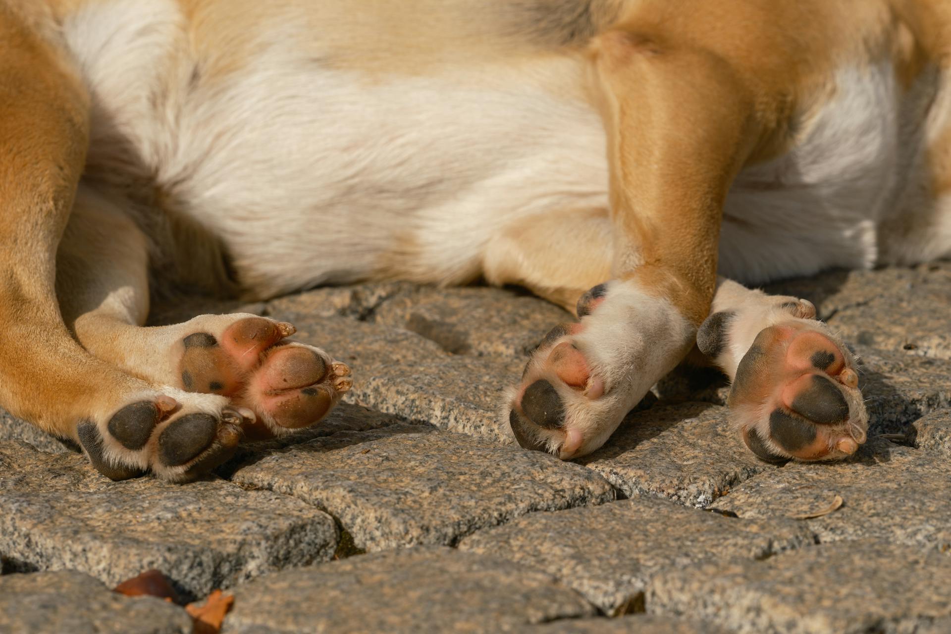
The mammary glands in dogs are located on the ventral aspect of the thoracic to inguinal regions, extending from the pectoral to inguinal regions.
You'll typically find paired mammary glands in female dogs, arranged in two rows bilaterally symmetric on the ventral aspect.
Mammary glands are usually arranged in two parallel rows extending from the underside of the chest to the groin area.
In dogs, there are usually five mammary glands on each side, joined together in a chain.
The mammary glands are located along the outside of the body wall, making them easily accessible for examination or care.
Here's an interesting read: Canine Eye Anatomy
Development of Mammae
In the embryonic development period, a mammary ridge is present in the fetus’s ventral aspect, which is responsible for the location of regular and supernumerary mammae in female dogs.
This mammary ridge extends from the axillary to the inguinal region of female dogs, and it's well developed in the dog compared to other mammals.
Take a look at this: Female Canine Reproductive Anatomy
In embryonic development, paired individual papillae mammae can be seen along the parental mammary ridge, typically arranged in 2 rows, but uneven numbers of mammae can occur in female dogs.
The most cranial thoracic mammary gland of the female dog may be absent in some cases, and the male fetus of the dog does not have the caudal inguinal mammary glands.
Mammary glands in dogs are usually arranged in two parallel rows extending from the underside of the chest to the groin area, along the outside of the body wall.
In dogs, there are usually five mammary glands on each side, joined together in a chain.
The development process of the dog mammary gland anatomy is complex, but understanding the embryonic development of the mammary ridge provides a solid foundation for understanding the anatomy of the mammary glands in dogs.
Intriguing read: Anatomy of a Dogs Ear
Arteries of Thoracic
The thoracic mammary glands of female dogs receive their arterial supply from the cranial branches of internal thoracic arteries. These arteries penetrate the intercostal spaces of the dog's thorax.
Take a look at this: Canine Thoracic Limb Anatomy

The cranial and caudal pairs of dog mammary glands are also supplied by the intercostal and lateral thoracic arteries. This is in addition to the internal thoracic arteries.
The internal thoracic arteries are responsible for supplying the thoracic mammary glands. This is crucial for the health and function of the glands.
Here's a summary of the arterial supply to the thoracic mammary glands:
- Internal thoracic arteries (branches)
- Branches from intercostal and lateral thoracic arteries
Arterial Supply and Drainage
The arterial supply to a dog's mammary glands is quite fascinating. The main arteries that supply the mammary glands are the cranial branches of the internal thoracic arteries, intercostal and lateral thoracic arteries, mammary branches of superficial epigastric arteries, cranial superficial epigastric arteries, and branches from the external pudendal artery.
These arteries bring oxygenated blood to the mammary glands, which are highly vascular. In fact, you'll find more extensive veins compared to arteries in the dog's mammary gland anatomy.
Here are the main arteries that supply the dog's mammary glands:
- Cranial branches of the internal thoracic arteries
- Intercostal and lateral thoracic arteries
- Mammary branches of superficial epigastric arteries
- Cranial superficial epigastric arteries
- Branches from the external pudendal artery
It's worth noting that the branches of the cranial and caudal superficial epigastric veins are the prominent ones that drain blood from the dog's mammary glands.
Veins of the

The veins of the dog's mammary glands run parallel to the arteries, almost 90% of the time. This is a notable characteristic of the venous system in dogs.
The cranial and caudal superficial epigastric veins are prominent in draining blood from the dog's mammary glands. These veins play a crucial role in the venous drainage of the mammary glands.
The branches of the caudal superficial epigastric veins drain blood from the abdominal and inguinal mammary glands. This is an important aspect of the venous drainage system in dogs.
The cranial superficial epigastric veins, on the other hand, drain blood from the thoracic mammary glands of female dogs. This is a key function of the venous system in dogs.
The internal thoracic veins also drain blood from the thoracic mammary glands of dogs at the level of the fifth intercostal space. This is a notable feature of the venous drainage system in dogs.
Here is a summary of the main veins that drain blood from the dog's mammary glands:
- Cranial superficial epigastric veins: drain blood from thoracic mammary glands
- Caudal superficial epigastric veins: drain blood from abdominal and inguinal mammary glands
- Internal thoracic veins: drain blood from thoracic mammary glands at the level of the fifth intercostal space
Arterial Supply
The arterial supply to a dog's mammary glands is a complex network of blood vessels that provide oxygen and nutrients to the tissue. The main arteries responsible for supplying blood to the abdominal mammary glands are the cranial superficial epigastric artery and the caudal superficial epigastric artery, which anastomose with each other.
The cranial superficial epigastric artery arises from the cranial epigastric artery and penetrates the straight abdominal muscle of the female dog, sending mammary branches to the cranial abdominal mammae.
The mammary glands are highly vascular, with more extensive veins compared to arteries. The main arteries that supply the dog's mammae include the cranial branches of the internal thoracic arteries, intercostal and lateral thoracic arteries, mammary branches of superficial epigastric arteries, cranial superficial epigastric arteries, and branches from the external pudendal artery.
Here's a summary of the arterial supply to the different regions of the dog's mammary glands:
These arteries play a crucial role in maintaining the health and function of the mammary glands, and any disruption to this network can have serious consequences for the dog's overall well-being.
Canine Lymphatic Drainage
Canine Lymphatic Drainage is a complex system that plays a crucial role in the overall health of a dog. The lymphatic channels of the canine mammary gland vary on each side, with each gland having its own plexus of lymphatic channels.
These channels anastomose and encircle the base of the papillary part of the dog mammary glands. The lymphatic channels are also distributed in the parenchyma and subcutaneous tissue of the dog mammae.
One to three main channels from each gland leave and pass superficially to the nearest lymph nodes. The thoracic mammary glands drain lymph directly to the axillary lymph nodes.
The caudal abdominal mammary glands drain lymph in two ways. The inguinal mammary glands have an extensive interlocking lymphatic plexus that drains the lymph into the adjacent superficial inguinal lymph nodes.
Understanding canine lymphatic drainage can help identify potential issues and ensure proper care for our furry friends.
How Many Glands in Mammals?
Dogs typically have 5 pairs of mammary glands on the ventral aspect, but you may also find 4 or 6 pairs.
Some dogs have four pairs of mammary glands, but this is quite rare.
Mammary glands in dogs arrange into 2 rows on the ventral aspect.
In the pectoral or thoracic region, you will find 2 pairs (4) of mammary glands.
The abdominal region of the dog contains 2 pairs (4) mammary glands.
Only one pair of mammary glands is found on the ventral aspect of the dog's inguinal region.
Frequently Asked Questions
You should know the details of dog mammae's shape, size, numbers, location, and internal and external features. The dog mammary glands are located on either side of the body, from the armpits to the groin area.
Dog anatomy learners often ask how many mammary glands dogs have. Dogs have 10 mammary glands, five on each side of the body.
The details of dog mammae's shape and size are crucial to understanding their anatomy. Dog mammary glands vary in shape and size, but they are generally teat-like in shape.
Dogs have external features like nipples and teats, but what about their internal features? The internal features of dog mammary glands are not as well-known, but they are essential for understanding the anatomy of the mammary glands.
A good understanding of dog mammary gland anatomy requires knowledge of their location on the body. The mammary glands are located on either side of the body, from the armpits to the groin area.
Dog anatomy learners often ask about the internal and external features of dog mammary glands. The internal features include the mammary glands themselves, while the external features include nipples and teats.
For your interest: Canine External Anatomy
Frequently Asked Questions
What does a swollen mammary gland look like on a dog?
A swollen mammary gland on a dog may appear red, purple, or discolored, with visible open wounds and scabs. If you suspect mastitis in your dog, consult a veterinarian for proper diagnosis and treatment.
Sources
- https://anatomylearner.com/dog-mammary-gland-anatomy/
- https://www.ncbi.nlm.nih.gov/pmc/articles/PMC7158309/
- https://www.ivis.org/library/mechanisms-of-disease-small-animal-surgery-3rd-ed/mammary-gland-disorders-of-dog-and-cat
- https://en.wikivet.net/Mammary_Gland_-_Anatomy_%26_Physiology
- https://www.petplace.com/article/dogs/pet-health/structure-and-function-of-the-mammary-system-in-dogs
Featured Images: pexels.com