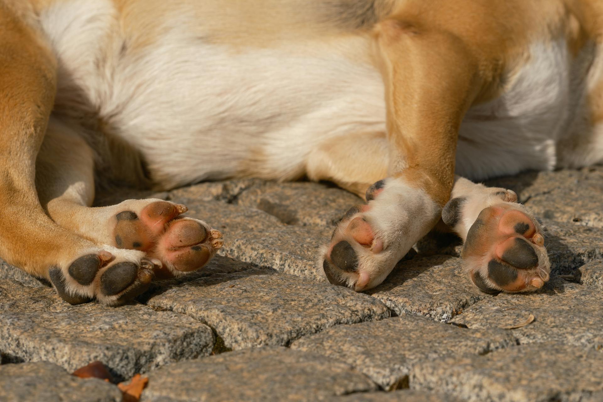
The canine pelvic limb is a remarkable structure that plays a crucial role in a dog's mobility and overall health. The pelvic limb is composed of the femur, patella, tibia, fibula, and metatarsal bones, which work together to support the dog's body weight and facilitate movement.
The femur, or thighbone, is the longest and strongest bone in the pelvic limb, accounting for about 70% of the limb's length. It connects the hip joint to the knee joint, enabling the dog to move its leg in a wide range of motion.
The patella, or kneecap, is a small, triangular bone that sits on top of the femur and helps to stabilize the knee joint during movement. The tibia and fibula, the two bones of the lower leg, work together to support the dog's body weight and transfer forces from the femur to the metatarsal bones.
The metatarsal bones, also known as the long bones of the foot, are responsible for bearing the dog's body weight and facilitating movement. They are connected to the phalanges, or toe bones, which help to distribute the dog's weight evenly across the ground.
Discover more: Canine Tibia Anatomy
Muscle Anatomy
The pelvic limb of a racing greyhound is made up of 21 muscles, which account for 18.5% of its total body mass. This is a significant proportion, considering the limb's importance in movement and locomotion.
The heaviest muscle in the pelvic limb is the biceps femoris, weighing in at 485 ± 58 grams. This is a substantial amount, especially considering it constitutes 18% of the total muscle mass of the limb.
Muscle mass decreases as you move down the limb, with the smallest muscles found in the distal (farthest from the body) part of the limb. Conversely, the largest muscles are located in the proximal (nearest to the body) part of the limb.
The gastrocnemius, flexor digitorum profundus, and flexor digitorum superficialis muscles have the smallest architectural indices (AIs), ranging from 0.07 to 0.14. This indicates a relatively low proportion of muscle fibers to total muscle mass in these muscles.
On the other hand, the semitendinosus, semimembranosus, and sartorius muscles have the largest AIs, ranging from 0.70 to 0.88. This suggests a higher proportion of muscle fibers to total muscle mass in these muscles, making them more efficient for movement.
Consider reading: Canine Thoracic Limb Anatomy
Joint Moments

At the hip and tarsus joints, the potential extensor moment is higher than the potential flexor moment. This is not the case at the stifle joint, which may reflect the presence of a large number of biarticular hip and tarsus extensor muscles.
The highest moment generating capacity is in the hip extensor musculature, which is consistent with the commonly held view that quadrupeds power locomotion by a torque about the hip joint. This is created by the hip extensor musculature, such as semitendinosus and semimembranosus.
The ratio of muscle moment arm to muscle fascicle length provides information regarding the roles of muscles in movement, and corroborates the view that hip extensors are key to locomotion.
Broaden your view: Canine Anatomy Muscles
Muscle Moment Arms
The muscle moment arms of the major pelvic limb muscles at the hip, stifle, and tarsus joints were obtained using the tendon travel method. This method measures the distance a tendon moves while the limb travels through an angle in radians.
On a similar theme: Canine Limb Anatomy
The moment arm of biceps femoris at the hip remained constant with changing joint angle. Semimembranosus showed a small increase in moment arm as joint angle increased.
At the stifle joint, moment arms of semitendinosus, gracilis, gastrocnemius, and biceps femoris all decreased linearly with increasing stifle extension. The mean maximum moment arm was greatest in semitendinosus at maximum joint flexion.
The total maximum torque that the combined pelvic limb musculature can produce at each joint is higher in the hip extensor musculature. This is consistent with the commonly held view that quadrupeds power locomotion by a torque about the hip joint.
Muscle anatomy and architecture play a crucial role in determining muscle moment arms. The mean total mass of the major muscles of the pelvic limb was 2769 ± 46 g, with biceps femoris being the heaviest muscle within the limb.
The ratio of muscle moment arm to muscle fascicle length gives us information regarding the roles of muscles in movement. This ratio distinguishes which muscles operate over a wide range of motion and which ones are more suited to 'holding' or maintaining posture.
Consider reading: Canine Stifle Joint Anatomy
Dog ACL
Dog ACL is actually a misnomer when it comes to our furry friends. Dog's technically do not have an ACL, which stands for the anterior cruciate ligament in humans.
The CCL, or cranial cruciate ligament, is a knee (or stifle) ligament in dogs that's prone to tears. Torn CCLs are a common rear leg dog injury.
Ligaments stabilize joints, and ligament injuries are fairly common in both humans and dogs.
Significance and Measurements
Understanding the significance of canine pelvic limb anatomy requires a grasp of its key measurements.
The canine pelvic limb is made up of the femur, patella, tibia, fibula, and metatarsals, which are connected by joints that allow for movement.
A typical dog's pelvic limb spans about 1/3 of their body length from the base of the neck to the tip of the tail. The femur, or thigh bone, is the longest bone in the pelvic limb, measuring around 4-6 inches in length.
See what others are reading: Canine Femur Anatomy
The patella, or kneecap, is located in the stifle joint, which is a modified hinge joint that allows for flexion and extension. The tibia and fibula, the two bones of the lower leg, are connected by the interosseous membrane.
The metatarsals, the long bones of the foot, are connected to the phalanges, or toe bones, which support the dog's weight and facilitate movement.
Anatomy of Specific Joints
The hip joint is a crucial part of a dog's pelvic limb anatomy, and it's responsible for generating a lot of power during locomotion. The hip extensor musculature, particularly the semitendinosus and semimembranosus muscles, are capable of producing high hip torques at various limb postures.
The stifle joint, on the other hand, has a unique characteristic where the potential extensor moment is lower than the potential flexor moment, which may be due to the presence of biarticular hip and tarsus extensor muscles.
The hock, also known as the dog ankle, connects the shin bones to the paw bones and is an essential part of a dog's hind leg anatomy.
Dog Knee
The dog knee, also known as the stifle joint, is a complex and delicate part of a dog's anatomy.
The stifle joint connects the femur, or thigh bone, to the tibia and fibula, the lower leg bones, and the patella, which is the canine equivalent of the knee cap.
Dogs often suffer from stifle joint-related injuries, including torn ACLs, which are technically called CCLs, or cranial cruciate ligaments.
Torn CCLs are a common rear leg dog injury, and it's essential to learn more about dog CCL tears and treatment options.
The stifle joint is a critical part of a dog's mobility, and injuries to this area can cause significant pain and discomfort.
The technical term for a dog knee is the stifle joint, and it's located on the hind legs.
Patellar luxation occurs when the dog's kneecap is dislocated from its normal position, which can be a painful and debilitating condition.
A dog's knee is made up of several bones, including the femur, tibia, fibula, and patella, all of which work together to provide support and mobility.
For your interest: Canine Rear Leg Anatomy
Dog Ankle
The dog ankle, also known as the hock, is a crucial part of a dog's hind leg anatomy.
It connects the shin bones to the paw bones, forming a vital joint that allows dogs to run, jump, and play.
A dog's hock is similar in function to the human ankle, but it's designed to support the unique weight and movement patterns of a dog's body.
Injuries to the hock can be painful and debilitating for dogs, which is why it's essential to learn more about canine hock injuries and how to prevent them.
By understanding the anatomy and function of the hock, dog owners can take steps to protect their furry friends from potential harm.
Fig. 6
Fig. 6 is a fascinating illustration that helps us understand the anatomy of specific joints, particularly in the context of muscle power output.
The schematic representation (A) shows how increased muscle volume and Architectural Index can lead to higher muscle power output, with powerful muscles residing in the pale blue region.
Muscle power output is closely related to muscle volume, but it's essential to consider other factors like fibre type compositions and contraction velocities, which can differ between breeds, species, and individual muscles.
The greyhound's pelvic limb muscles are capable of generating substantial amounts of work, making them ideal for high-speed locomotion.
Biceps femoris and adductor muscles are the largest and most powerful hip extensors, while rectus femoris is particularly powerful due to its relatively short fibres.
Rectus femoris has sufficient power and force-generating capabilities to be a potential candidate for creating rapid hind limb protraction.
Tables and Data
In canine pelvic limb anatomy, understanding the physical characteristics of dogs is crucial. The pelvic limb segment lengths used to scale muscle moment arm measurements are an essential aspect of this study.
The data in Table 3 shows the femur length of dog number 1 is 19.0 cm, while dog number 2 has a femur length of 20.0 cm. A 1 cm difference in femur length can significantly impact the muscle moment arm measurements.
The mass of the dogs in the study ranges from 27 kg to 34 kg, with dog number 1 weighing 27 kg and dog number 2 weighing 34 kg. This variation in mass is another important factor to consider when scaling muscle moment arm measurements.
Table 2
Table 2 is a treasure trove of information about the pelvic limb tendons in the human body. It provides data on the mass, volume, and resting length of these tendons, which is crucial for understanding their function and behavior.
The tendon data in Table 2 includes the mass of the Semitendinosus tendon, which is 1.0 grams on average. This is a relatively small tendon, but it plays a significant role in the movement of the leg.
One of the most interesting aspects of Table 2 is the estimated cross-sectional area (CSA) of the tendons. The CSA of the Gracilis tendon is 5.4 square centimeters on average, which is relatively small compared to other tendons in the table.
The stress and strain of the tendons are also important factors to consider. The Semitendinosus tendon has a stress of 1.5 MPa on average, which is relatively low compared to other tendons in the table.
Additional reading: Why Are Chihuahuas so Small
Here's a summary of the tendons in Table 2, grouped by their CSA:
The length change of the tendons is also an important factor to consider. The Semitendinosus tendon has a length change of 15 millimeters on average, which is relatively small compared to other tendons in the table.
Table 3
In Table 3, we have a list of dog characteristics that are used to scale muscle moment arm measurements.
The table includes four different dogs, with varying sex, mass, femur length, and tibia length.
Dog number 1 is a female dog with a mass of 27 kg.
The femur length of dog number 1 is 19.0 cm.
The tibia length of dog number 1 is 23.0 cm.
Dog number 2 is a male dog with a mass of 34 kg.
The femur length of dog number 2 is 20.0 cm.
The tibia length of dog number 2 is 27.0 cm.
Dog number 3 is a male dog with a mass of 33 kg.
The femur length of dog number 3 is 20.5 cm.
The tibia length of dog number 3 is 27.0 cm.
Dog number 4 is a male dog with a mass of 28 kg.
The femur length of dog number 4 is 20.0 cm.
The tibia length of dog number 4 is 26.0 cm.
Here's a summary of the dog characteristics in Table 3:
Sources
- https://www.ncbi.nlm.nih.gov/pmc/articles/PMC2644771/
- https://vanat.ahc.umn.edu/carnLabs/Lab05/Lab05.html
- https://www.cram.com/flashcards/canine-pelvic-limb-anatomy-2330927
- https://www.semanticscholar.org/paper/Two-anatomic-resources-of-canine-pelvic-limb-based-Sunico-Hamel/42a2578d16347cd1b1f9fd055a7f10408790ec12
- https://orthodog.com/article/dog-leg-anatomy/
Featured Images: pexels.com