Canine skulls come in a wide range of shapes and sizes, reflecting the diversity of breeds and their unique characteristics. The skull is made up of several bones that fuse together as the dog matures.
The cranium, which forms the upper part of the skull, is a key feature that distinguishes one breed from another. For example, the cranium of a Greyhound is long and narrow, while that of a Bulldog is short and broad.
The muzzle, which forms the lower part of the skull, varies greatly in length and shape among breeds. Some breeds, like the Pug, have a short, compact muzzle, while others, like the Whippet, have a long, narrow one.
The jawbone, or mandible, is another important feature of the canine skull. In some breeds, like the German Shepherd, the jawbone is long and narrow, allowing for a powerful bite, while in others, like the Pekingese, it is shorter and more compact.
Intriguing read: Rear Dew Claws on Dogs Breeds
Bones of the Canine Skull
The occipital bone is the most caudal bone of the canine skull, forming the external occipital protuberance which provides attachment for the nuchal ligament.
This bone has nuchal crests laterally and a sagittal crest medially and dorsally to the external occipital protuberance. The sagittal crest is often prominent and can be palpated in most canines unless they are very well muscled.
The occipital bone also has muscular tubercules on its ventral surface where the flexors of the head and neck attach, and a caudocranial fossa that encloses the pons and medulla oblongata.
For your interest: Canine External Anatomy
Occipital Bone
The occipital bone is the most caudal bone of the skull. It's a vital part of the canine skull, forming the back and base of the cranium.
The external occipital protuberance, located medially within the bone, provides the attachment for the nuchal ligament. This ligament plays a crucial role in supporting the head and neck.
The occipital bone has nuchal crests laterally and a sagittal crest medially and dorsally to the external occipital protuberance. The sagittal crest is prominent and can be palpated in most canines.
The pars basilaris element is the caudal base of the cranium, although rostral to foramen magnum and joined by a cartilagenous suture to basisphenoid bone. This element is crucial for the attachment of muscles that flex the head and neck.
The squamous part of the occipital bone, or pars squamosa, is dorsal to the lateral parts and occipital condyles. The occipital condyles articulate with the atlas to form the atlanto-occipital joint, allowing for flexibility and movement of the head.
The paracondylar process provides muscle attachment sites for muscles of the head. This is an important feature for canine anatomy, as it enables the head to move and flex in various ways.
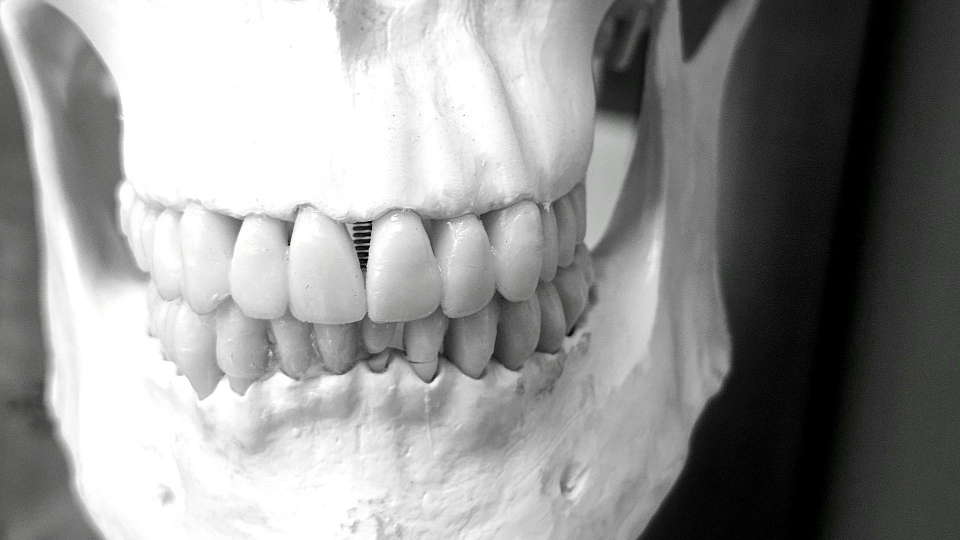
Mandible (Mandibula)
The mandible, also known as the mandibula, is a crucial bone in the canine skull that plays a key role in supporting the teeth.
It can be divided into two main parts: the body and the ramus. The body of the mandible supports the incisor teeth rostrally and the cheek teeth caudally.
Explore further: Types of Dog Teeth
The section of the body without teeth is called the interalveolar margin or diastema. This area is quite distinct from the rest of the body.
The mandible also contains the mandibular canal and the mental foramen. These features are essential for the proper functioning of the bone.
The facial notch is located on the ventral surface of the mandible, where the facial vessels run. This notch is a vital passageway for these vessels.
The ramus extends from the caudal end of the body dorsally towards the zygomatic arch. This extension provides a solid base for the attachment of muscles.
The masseter muscle attaches to the lateral surface at the masseteric fossa. This attachment point is strong and secure.
The angle of the mandible terminates dorsally in the condylar process and the coronoid process, which are separated by the mandibular notch. This unique arrangement allows for flexibility and movement.
The temporal muscle inserts onto the coronoid head. This insertion point is essential for the proper functioning of the muscle.
The condylar process articulates with the mandibular process of the skull. This articulation is a critical connection between the mandible and the skull.
You might enjoy: Dogs Muscle Anatomy
Skull Anatomy and Variation
Skull shape has varied significantly since dog domestication began. Molecular clock estimates suggest this started as early as 135,000 years ago.
Prehistoric dog skulls excavated in Russia were from massive animals with shortened snouts and widened palates. This is a clear example of how skull shape has changed over time.
The overall size of dogs' brains relative to wolves has decreased by nearly 30%. This is particularly acute in the limbic system, which is integral to fight or flight responses.
In contrast, modern dogs exhibit an increased brain-to-body-size ratio, a trait shared with other large domesticates.
Skull Shape and Domestication Variation
Skull shape has changed significantly in dogs since their domestication. Domestication started as early as 135,000 years ago, with more conservative estimates ranging from 15,000 to 36,000 years ago.
The skull was at the leading edge of anatomical changes in incipient dogs, with prehistoric dog skulls found in Russia having shortened snouts and widened palates. In contrast, ancient dogs were often smaller than wolves.
Modern dogs exhibit an increased brain-to-body-size ratio, but their overall brain size relative to wolves has decreased by nearly 30%. This decrease is particularly notable in the limbic system, which is integral to fight or flight responses.
Domestication may have reduced areas of the wolf brain that enabled tolerance to human contact. As a result, skull shape likely changed in response to changes in brain morphology.
Some researchers have likened dogs to wolf pedomorphs, proposing that dogs are juvenilized wolves that are developmentally restrained in behavior and physical maturation.
You might enjoy: Canine Brain Anatomy
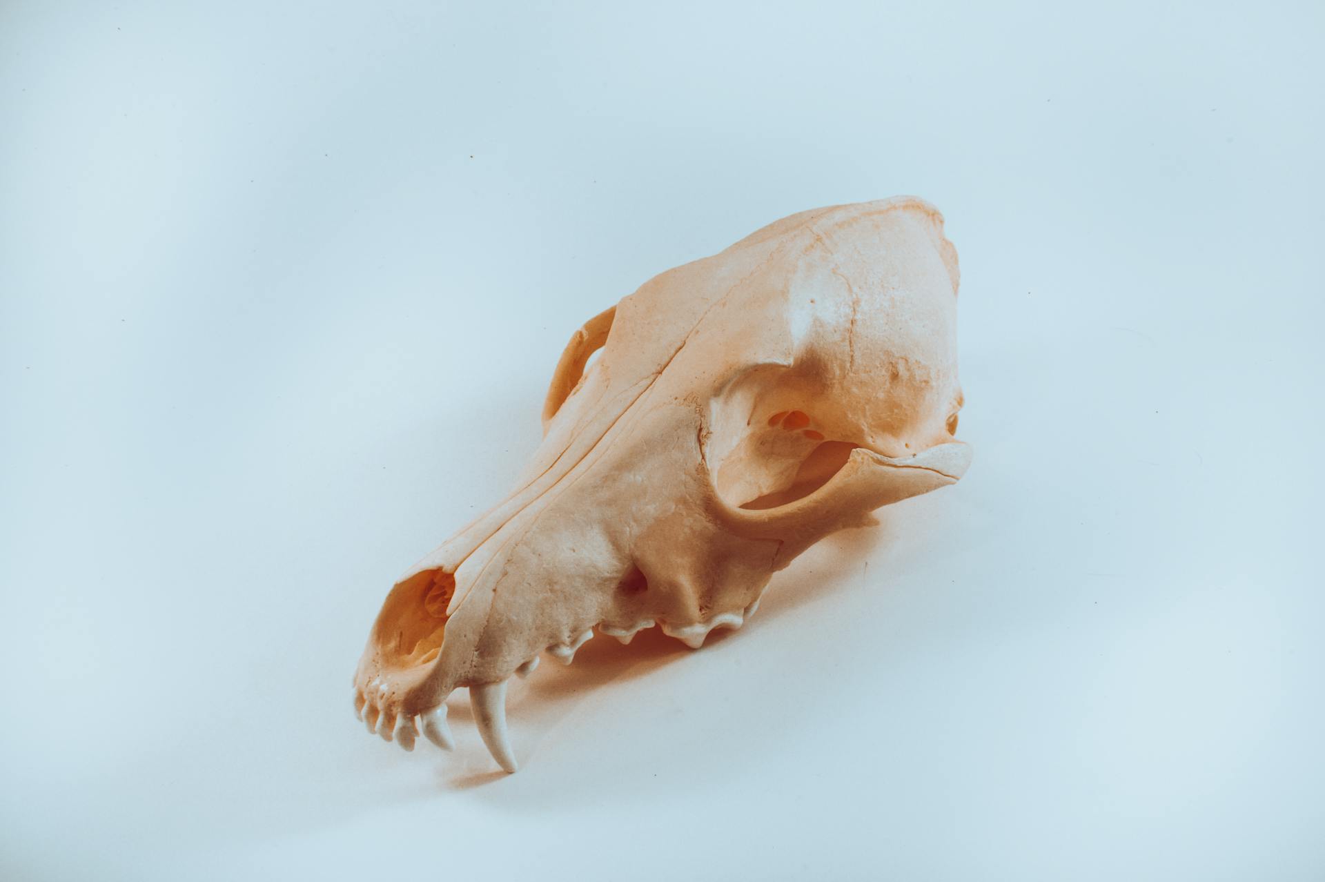
Dog Skull (Large)
The dog skull is a fascinating specimen that offers a unique opportunity to study the anatomy of Canis domesticus. This real canine skull has flexible mounting to enable natural movement of the jaw, making it an excellent educational aid for comparative veterinary anatomy study.
The dog skull model represents a large-sized dog, with a skull length of approximately 22.5 cm. This makes it a great reference point for understanding the anatomy of larger breeds.
Our real animal skeleton range, prepared by professional taxidermists in Europe, ensures that every bone from the animal is displayed to the highest quality. No animals have been bred or killed solely for the purpose of making these specimens, and these models are certified to pose no risk of infection due to infectious zoonotic pathogens.
Genetic Factors
Genetic Factors play a significant role in shaping a dog's skull anatomy.
The shape and size of a dog's skull are determined by the interaction of multiple genes, which can vary between breeds.
For example, the brachycephalic breeds such as Pugs and Bulldogs have a short, compact skull due to their genetic makeup.
Emergence of Dog Breeds
The emergence of dog breeds is a fascinating story that began in the 19th century in Europe. By the 1870s, dog fanciers were actively promoting and breeding dogs with specialized physical and behavioral traits.
Dog fanciers quickly recognized that structured breeding could be used to transmit desirable traits, but they didn't fully understand the genetic mechanisms behind it. It wasn't until Charles Stockard conducted detailed studies on dog pedigrees that the complexity of breed-defining traits became clear.
Stockard's research showed that breed-defining traits, such as the bulldog's shortened rostrum, didn't follow patterns of Mendelian inheritance. This was a crucial discovery that would take nearly 70 years to be confirmed by genome-wide association studies.
The acceptance of the dog as a system for studying genetics was key to advancing our understanding of mammalian developmental biology. With over 400 documented breeds worldwide, geneticists could study complex traits and their genetic underpinnings in a way that was not possible in human populations.
The development of genetic and physical maps of the dog genome made it a valuable model organism for genetic studies. By 2005, a 7.5× sequence and draft assembly of the dog genome was completed, putting it on par with traditional model organisms like mice and humans.
Genetic Factors
Genetic Factors play a significant role in determining our susceptibility to various diseases.
Research has identified over 1,000 genetic variants associated with an increased risk of developing type 2 diabetes. The most significant risk factor is having a family history of the disease, with a 50% increase in risk for those with a first-degree relative affected.
Genetic testing can identify specific genetic variants that increase the risk of developing certain diseases, such as cystic fibrosis. This can help families make informed decisions about their reproductive choices.
Many genetic disorders, such as sickle cell anemia, are caused by a single genetic mutation. This mutation can be inherited from one's parents, highlighting the importance of genetic counseling for families with a history of these disorders.
Genetic factors can also influence an individual's response to certain medications, such as warfarin, which requires regular genetic testing to ensure safe dosing.
Canine Skull Images
Check out these incredible canine skull images. A montage of canine craniofacial shape demonstrates the incredible morphologic diversity of Canis familiaris.
The images show various breeds of dogs from different perspectives, including dorsal, lateral, and ventral views. The lateral views are articulated so that the skull base is approximately parallel between breeds.
You can see prominent differences across breeds, including palate shape, neurocranium shape, and cranial base length. The angle of the palate relative to the cranial base also varies between breeds.
Figure 1
Looking at Figure 1, you can see the incredible diversity of canine craniofacial shape. A montage of various dog breeds showcases their unique morphologic features.
The lateral views of different breeds have their skull bases (red line) approximately parallel to each other, which helps to compare their features. This allows us to see the differences in palate shape and neurocranium shape across breeds.
Dorsal, lateral, and ventral perspectives of the breeds are shown, highlighting their distinct characteristics. The palate shape and neurocranium shape are particularly prominent in this figure.
Prominent differences across breeds include palate shape, neurocranium shape, and cranial base length. These features are crucial for breed identity and domestication.
The angle of the palate relative to the cranial base is also notable in this figure. This angle is an important aspect of canine skull morphology.
Figure 2
Canine skull length is a complex trait, with different breeds exhibiting unique characteristics. Brachycephalic dog breeds have a shortened rostrum, wide zygomatic arches, and a rounded neurocranium.
The surface scans of a wolf skull morphed to illustrate the differences between brachycephalic, ancestral, and dolichocephalic skull states of canids are quite striking. Brachycephalic breeds have a distinct appearance that sets them apart from other breeds.
In contrast, dolichocephalic dog breeds have a narrow, sometimes elongated, snout and orbitals that are less forward set. These breeds include the Saluki, Borzoi, and collie.
Dolichocephalic breeds were originally bred for coursing small prey, and their craniofacial configuration is well-suited for this purpose. A dolichocephalic morphology is exactly what one would predict based on the relationship between morphology and ecology/hunting behavior of wild canids.
Angulation between the skull base and hard palate also varies significantly between dog breeds. Klinorhynchy, the hallmark downward-pointing snout of bull terriers, is a notable example of this variation.
Figure 3
Figure 3 shows the craniofacial diversity that exists between and within breed dogs, with white strips highlighting the palate and brainstem in each skull example.
The continuum of airorhynchic and klinorhynchic dog breeds is arranged in order of severity, featuring breeds like the Pekingese, French bulldog, Chow Chow, Bernese Mountain Dog, German Shepherd, and Borzoi.
These breeds demonstrate a range of skull shapes and sizes, from the short, flat face of the Pekingese to the longer, more pointed face of the Borzoi.
Bull terrier skulls, on the other hand, show a continual morphological evolution in breed dogs, with skulls arranged chronologically from the oldest to the most modern.
The oldest bull terrier skulls have a more traditional, longer face, while the most modern skulls have a shorter, more compact face.
Materials and Methods
To study canine skull anatomy, we began by collecting 20 canine skulls for our sample.
We used a flowchart to represent the simplified procedure for a single canine skull, which was repeated for each of the 20 skulls in the sample.
This procedure allowed us to calculate the mean errors for each of the four types of CT models compared to their corresponding laser scans.
Each CT model was compared to its corresponding laser scan to determine the accuracy of the digital representation.
The 20 mean errors calculated for each CT model type provided a comprehensive understanding of their precision.
Frequently Asked Questions
What is the bony protrusion on a dog's head?
The bony protrusion on a dog's head is called an "occiput." It serves as a natural protective feature for the dog's skull and brain.
What are the parts of a dog's head?
A dog's head consists of the nose, muzzle, stop, forehead, occiput, ears, eyes, eyebrows, whiskers, flews, and cheeks. These distinct features work together to create a unique and recognizable canine face.
What is the indentation in the dog's forehead called?
The indentation in a dog's forehead is called the stop. It varies in depth among different breeds.
Featured Images: pexels.com